
Definition
• An incidental thymic involution in response to various stressful conditions; different from slow physiologic age-induced thymic involution
Pathogenesis
• In stressful events, sudden secretion of corticosteroid by the adrenal cortex causes rapid depletion of thymic cortical lymphocytes (thymocytolytic effect)
Clinical features
Epidemiology
• Usually related to stress or infection
• Seen in newborn infants with chorioamnionitis and sepsis, especially in premature ones
• Also seen in patients with malnutrition, immunosuppression, or cancer chemotherapy
Presentation
• Acute involution is seen at autopsy of neonates and preterm infants; no specific clinical symptoms during life
• Adults with acute thymic involution usually present with decreased T-cell mediated immunity
Prognosis and treatment
• Treat underlying acute illness; steroid inhibitor RU486 has been tried in a mouse model to reverse thymic involution
• Prognosis depends on duration of underlying illness: short-term involution is reversible; prolonged involution may destroy the thymus
Pathology
Histology
• Histologically graded (0-4) by van Baarlen method:
• Grade 0: resting state with normal thymic architecture
• Grade 1: mild cortical lymphophagocytosis with scattered large pale macrophages
• Grade 2: prominent cortical lymphophagocytosis with a moth-eaten, starry-sky appearance; Hassall corpuscles may be necrotic
• Grade 3: loss of sharp corticomedullary demarcation or even reversed corticomedullary cellularity with an inside-out appearance; Hassall corpuscles become prominent and appear right beneath the capsule
• Grade 4: advanced lymphodepletion and relative prominence of perivascular spaces; Hassall corpuscles may disappear and are replaced with foamy macrophages
• The grade of acute thymus involution correlates with the duration and severity of acute illness in neonates
Main differential diagnoses
• Thymic dysplasia:
• Present in congenital inherited immunodeficiency syndrome
• An abnormality of thymic epithelial development with near absence of Hassall corpuscles and associated with thymocyte depletion; may mimic severe involution
• Accompanied by poorly developed lymph nodes and spleen

Fig 1 Acute involution of thymus. Grade 1 acute thymic involution: low (A) and medium (B) powers; note the scattered clear spaces with large pale macrophages.
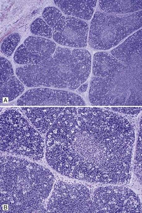
Fig 2 Acute involution of thymus. Grade 2 acute thymic involution: low (A) and medium (B) powers; note moth-eaten, starry-sky appearance.

Fig 3 Acute involution of thymus. Grade 3 acute thymic involution: low (A), medium (B), and high (C) powers. Note pronounced cortical lymphodepletion with loss of normal corticomedullary distinction, prominent perivascular spaces, and Hassall corpuscles.
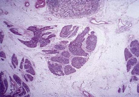
Fig 4 Acute involution of thymus. Grade 4 acute thymic involution with small lobules widely separated by fibrous tissue and almost complete depletion of lymphocytes.