
Definition
• Enlarged thymus (weight greater than upper limit of normal for age) with normal histological appearance
Clinical features
Epidemiology
• The most common cause of an anterior mediastinal mass in infants and children
• Can occur in adults, often after stress such as chemotherapy, severe burns, irradiation, or steroid therapy (referred to as rebound hyperplasia)
Presentation
• Usually asymptomatic and detected on chest x-ray films
• Rarely, can have massive thymic hyperplasia causing compression of surrounding structures and resulting in respiratory symptoms and/or infection
• Can be associated with autoimmune diseases, myasthenia gravis, or hyperthyroidism
Prognosis and treatment
• May regress spontaneously or with steroid treatment
• If persistent, symptomatic, or calcified, complete excision to relieve symptoms and confirm diagnosis is warranted
Pathology
Gross
• Increased size and weight (as compared with age-matched control subjects)
Histology
• Normal thymic tissue
• Lobular architecture and corticomedullary junction are preserved
Immunopathology/special stains
• Not contributory
Main differential diagnoses
• Lymphoid hyperplasia of thymus: usually of normal size and weight; lymphoid follicles with germinal centers are seen on histological analysis
• Thymoma: normal architecture effaced
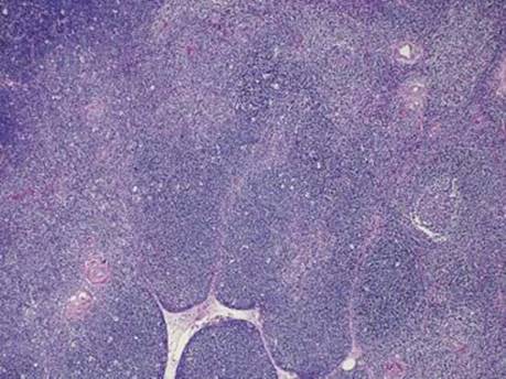
Fig 1 True thymic hyperplasia. Low power demonstrating normal lobular architecture of the thymus.
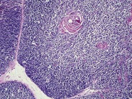
Fig 2 True thymic hyperplasia. Note the maintained lobular configuration and preserved corticomedullary junction (medium power).
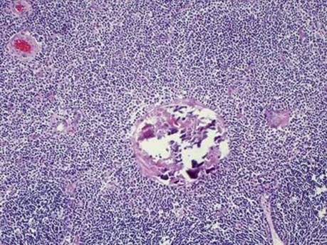
Fig 3 True thymic hyperplasia. High-power view of a calcified Hassall corpuscle.