
Definition
• An intermediate-grade thymoma composed of sheets of medium-sized polygonal epithelial cells with slight atypia and few or no intermixed lymphocytes
Clinical features
Epidemiology
• Accounts for <25% of all thymomas
• Mean age, 45 to 50 years
• No gender predilection
Presentation
• Myasthenia gravis is the most common presentation
• Frequent local symptoms like chest pain, coughing, or shortness of breath
Prognosis and treatment
• Usually unresectable tumor; treated with chemotherapy and radiation therapy
• Intermediate malignancy with frequent local recurrence and metastasis; more aggressive than type B2 thymoma with 10-year survival rate of 50% to 70%
Pathology
Gross
• Usually not encapsulated; extension into mediastinal fat or adjacent organs
• Cut surface is firm with white-tan nodules separated by white fibrous bands
• May contain areas of cystic changes or calcification
Histology
• Sheets or nests of confluent tumor cells with lobular growth pattern separated by thick fibrous septa
• Intraepithelial lymphocytes are scattered or absent
• Tumor cells are medium-sized polygonal cells with round to elongated, irregular nuclei; they are smaller than in type B2 thymoma and have less prominent nucleoli but more atypia
• Overt palisading around perivascular spaces or along septae
Immunopathology/special stains
• Cytokeratin stains show confluent tumor epithelial cells
• Tumor epithelial cells are usually negative for CD5 and CD70
Main differential diagnoses
• B2 thymoma:
• More intraepithelial lymphocytes and less epithelial cells
• Epithelial cells are large and without atypia
• Low-grade squamous cell carcinoma:
• Lack organotypic pattern of conventional thymoma
• Keratinization and/or intercellular bridges are present
• Type A thymoma:
• Bland spindle cells with minimal or no atypia
• No perivascular or septal palisading

Fig 1 Type B3 thymoma. Type B3 thymoma with confluent tumor epithelial cells and few intermixed lymphocytes: low (A), medium (B), and high (C) powers; note normal thymus on the right in A and mild atypia and mitotic figures in C.
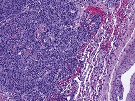
Fig 2 Type B3 thymoma. Type B3 thymoma invading the lung parenchyma (Masaoka stage III).

Fig 3 Type B3 thymoma. Type B3 thymoma with vascular invasion.

Fig 4 Type B3 thymoma. Immunohistochemical staining of type B3 thymoma: keratin AE1/AE3 stains the epithelial cells (A) and CD5 stains only lymphocytes (B).