
Definition
• An intermediate grade of thymoma composed of large polygonal epithelial cells intermixed with multiple background small lymphocytes
Clinical features
Epidemiology
• Most common or second most common subtype
• Mean age, approximately 50 years
• No gender predilection
Presentation
• More aggressive tumor than B1 thymoma; higher stage at initial presentation
• Often invasive; 5% to 15% of cases are not resectable at presentation
Prognosis and treatment
• Surgical resection; neoadjuvant therapy for nonresectable tumors
• Moderate malignancy with 10-year survival rate between 50% and 100% depending on resectability
Pathology
Gross
• Usually encapsulated or vaguely circumscribed; may invade mediastinal fat or adjacent organs
• Cut surface is soft or firm with white-tan nodules separated by white fibrous bands
• May have cystic changes and hemorrhage
Histology
• Tumor has a lobular growth pattern with delicate septa resembling normal thymic cortex
• Tumor is composed of large polygonal neoplastic epithelial cells with vesicular chromatin and prominent nucleoli, mixed with a large number of nonneoplastic immature T lymphocytes
• Neoplastic epithelial cells form a loose network and are arranged around perivascular spaces or along septae with a palisading appearance
• Lymphoid follicles are more frequently seen in myasthenia gravis–associated cases
• Combined B2/B3 thymoma:
• Occurs in a quarter of B2 thymomas
• Areas of B3 thymoma are composed of sheets of confluent neoplastic epithelial cell growth with few or no lymphocytes (lymphocyte-poor areas)
• Compared with B2 thymoma, the neoplastic epithelial cells are smaller with irregular nuclei and less prominent nucleoli
Immunopathology/special stains
• Cytokeratin highlights the loose network of tumor epithelial cells
• The intraepithelial lymphocytes have the staining pattern of cortical immature T lymphocytes
Main differential diagnoses
• Type B1 thymoma:
• Scattered epithelial cells with smaller size and inconspicuous nucleoli
• More abundant lymphocytes
• Type B3 thymoma:
• Almost no intermixed lymphocytes
• Confluent tumor epithelial cells with mild atypia
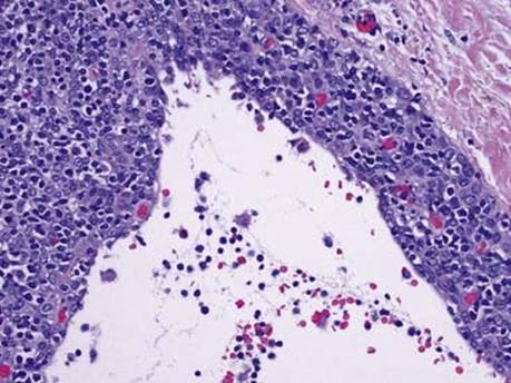
Fig 1 Type B2 thymoma. Type B2 thymoma with cyst formation.

Fig 2 Type B2 thymoma. Type B2 thymoma with large epithelioid tumor cells intermixed with multiple lymphocytes; low (A), medium (B), and high (C) powers.

Fig 3 Type B2 thymoma. Transbronchial needle aspirate of a mediastinal mass showing type B2 thymoma: low power (A), high power (B), and cytokeratin immunostain showing loose network of epithelial cells (C).