
Definition
• A tumor composed of myofibroblastic spindle cells with lymphoplasmacytic inflammation, associated with ALK gene translocations, previously referred to as inflammatory pseudotumor
Clinical features
Epidemiology
• Equal gender distribution
• Typically occurs in children but can be seen at any age
• IMT is the most common childhood mesenchymal endobronchial tumor but overall accounts for less than 1% of all lung tumors
• Some cases have been shown to be associated with a preceding human herpesvirus 8 infection
Presentation
• Peripheral tumors often asymptomatic and discovered incidentally on imaging
• Less commonly, endobronchial location, presenting with signs of airway obstruction, including coughing, wheezing, chest pain, and hemoptysis
• In many cases, patients have symptoms of an associated paraneoplastic syndrome with weight loss, fever, anemia, increased erythrocyte sedimentation rate, leukocytosis, and thrombocytosis, as well as hyperglobulinemia
Prognosis and treatment
• Complete excision usually leads to excellent survival rate
• Incomplete excision can lead to recurrence
• Extrapulmonary invasion and metastases are infrequent but are associated with a poor prognosis
Pathology
Histology
• Well-demarcated unencapsulated mass of varying size (average, 3 cm), which may focally extend beyond its circumscribed edges into the adjacent lung parenchyma
• Bland spindle cells growing in short intersecting fascicles
• Spindle cells have oval nuclei, occasional nucleoli, and abundant eosinophilic cytoplasm
• Mitotic figures are present in variable numbers, but there are no atypical mitotic figures
• Prominent inflammatory infiltrate of lymphocytes, plasma cells, and eosinophils, as well as clusters of foamy histiocytes and scattered Touton-like giant cells
• Stroma may be focally myxoid or fibrous and calcifications may be present
• Endobronchial tumors may have adjacent postobstructive pneumonia and atelectasis
Immunopathology/special stains
• Spindle cells are positive for vimentin, muscle-specific actin, calponin, smooth muscle actin, and occasionally desmin and ALK
• Spindle cells are negative for caldesmon, CD117, S100, CD34, EMA, myogenin, and myoglobin
• p53 may be positive in recurrence or malignant transformation
• Entrapped alveolar pneumocytes are cytokeratin positive
Main differential diagnoses
• Organizing pneumonia: look for characteristic Masson bodies
• Inflammatory sarcomatoid carcinoma: cytokeratin and EMA positive; muscle markers negative; also has more high-grade features: nuclear hyperchromasia, atypia, and necrosis
• Solitary fibrous tumor: CD34-positive spindle cells, less inflammation, fibrohyaline stroma
• Leiomyoma or leiomyomatous hamartoma: caldesmon positive, less inflammation
• Congenital peribronchial myofibroblastic tumor: rare, present at birth, extensively infiltrative with minimal inflammation
• Spindle cell sarcoma, NOS
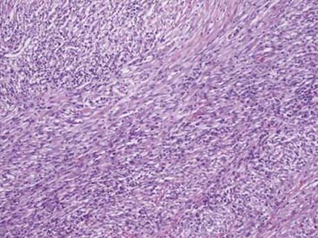
Fig 1 Inflammatory myofibroblastic tumor. Short intersecting fascicles of eosinophilic spindle cells admixed with inflammatory cells, as seen here, are characteristic of IMT.

Fig 2 Inflammatory myofibroblastic tumor. The fascicles of spindle cells vary from loose bluish (lower right) to fibrotic (upper left).

Fig 3 Inflammatory myofibroblastic tumor. The inflammatory infiltrate can be quite dense, as seen in this case of IMT.
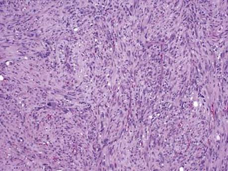
Fig 4 Inflammatory myofibroblastic tumor. At low power, this atypical IMT shows increased cellularity and cellular pleomorphism.
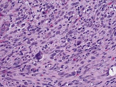
Fig 5 Inflammatory myofibroblastic tumor. At higher power, the atypical cells can be seen along with the more typical features of spindle and inflammatory cells.
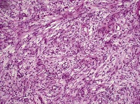
Fig 6 Inflammatory myofibroblastic tumor. On this frozen section of an IMT, spindle cells are seen admixed with lymphocytes and plasma cells.
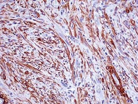
Fig 7 Inflammatory myofibroblastic tumor. Spindle cells are positive for SMA. Note positive staining for vascular smooth muscle at bottom right.
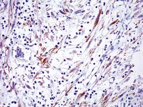
Fig 8 Inflammatory myofibroblastic tumor. ALK-1 shows both granular cytoplasmic and membranous staining.