
Definition
• A benign spindle cell tumor of the lung presenting at birth or in neonatal life with clinical features of respiratory distress and mediastinal shift
• It has also been termed congenital peribronchial myofibroblastic tumor
Clinical features
Epidemiology
• Rare neoplasm; less than 40 cases reported in the literature
Presentation
• Mass effect leads to respiratory distress
• Has been incidentally detected on prenatal ultrasonography
Prognosis and treatment
• It is a benign tumor that is cured with surgical resection
• Mortality is related to mediastinal shift (preoperatively) or due to complications of surgery (intraoperative and postoperative)
Pathology
Histology
• Well-circumscribed, usually nonencapsulated tumor with broad-pushing front; small foci of infiltrative growth may be present
• Interlacing fascicles of bland spindle cells of variable density; cells lack anaplasia
• Stroma may show myxoid change
• Foci of necrosis and dystrophic calcification may be present
• Mitotic activity varies from zero to four per 10 high-power fields
• Vascularity may be prominent focally and may show a hemangiopericytoma-like pattern
• Entrapped respiratory structures and cartilage usually present
Immunopathology/special stains
• Spindle cells positive for vimentin and may be positive for smooth muscle actin. Hence, in older literature, it was thought to represent a “congenital leiomyosarcoma”
• Desmin and S100 are negative
• Electron microscopy suggests myofibroblastic differentiation, in that the tumor cells have disrupted basal lamina, lack micropinocytotic vesicles, contain bundles of fine actin-like filaments and have crenated nuclei. Others have reported elongated cells with some cytoplasmic processes
Main differential diagnoses
• Sarcomas such as leiomyosarcoma (these will be desmin-positive) and malignant peripheral nerve sheath tumors (at least focally S100 positive). Sarcomas commonly show anaplasia, unlike congenital pulmonary myofibroblastic tumors
• Pleuropulmonary blastoma (PPB): types 1 and 2 PPB have a cystic component. Solid (type 3) PPB is sarcomatous and commonly shows features of a rhabdomyosarcoma

Fig 1 Congenital pulmonary myofibroblastic tumor. Low power shows nodules of tumor with irregular cartilage plates.
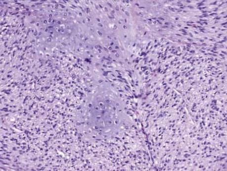
Fig 2 Congenital pulmonary myofibroblastic tumor. Intermediate power shows relatively bland tumor cells with islands of cartilage.
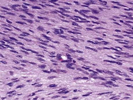
Fig 3 Congenital pulmonary myofibroblastic tumor. High power shows uniform spindle cells with one mitotic figure.