
Definition
• Uncommon cystic lesion of the posterior mediastinum usually lined by gastric epithelium
Pathogenesis
• Congenital foregut malformation
Clinical features
Epidemiology
• Usually discovered in neonates or infants
• Up to 30% are found in adults
• More frequent in males
Presentation
• Symptoms may include respiratory distress, dysphagia, coughing, recurrent pneumonia, chest pain, or hematemesis
• Complications can include peptic ulceration, perforation, and hemorrhage
• Often associated with vertebral malformations (especially cervical hemivertebra, spina bifida, vertebral fusion, scoliosis) and may be connected to vertebrae
Prognosis and treatment
• Treatment: surgical excision
• Usually benign lesions with excellent prognosis
• Can be life-threatening if peptic ulceration or perforation occurs
• Very rare reports of malignant transformation to adenocarcinoma or neuroendocrine carcinoma
Pathology
Gross
• Usually unilocular but can be multilocular
• Smooth lining may have areas of ulceration and hemorrhage
• Can be filled with serous, mucoid, or hemorrhagic fluid
• Located within esophageal wall or adherent to outer surface of esophagus
• Usually 2 to 10 cm but can measure up to 15 cm
Histology
• Lining of cyst is usually gastric epithelium but can be squamous, ciliated columnar, or intestinal-type epithelium
• Double layer of smooth muscle
• Often contains nerve fibers and ganglia
• May contain pancreatic tissue or an adrenal rest
Immunopathology/special stains
• Not contributory
Main differential diagnosis
• Bronchogenic cyst
• Esophageal cyst
• Cystic teratoma

Fig 1 Mediastinal gastroenteric cyst. High-power view of cyst lined by gastric epithelium.
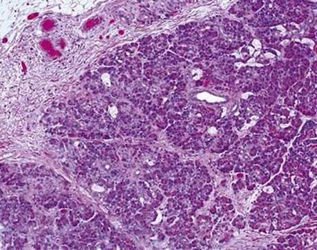
Fig 2 Mediastinal gastroenteric cyst. Gastroenteric cyst containing pancreatic tissue in the cyst wall.