
Definition
• Mesothelial-lined cyst attached to the pericardium
Pathogenesis
• Usually congenital but can be acquired
• Thought to be due to failure of all the lacunae that form the pericardial sac to merge
Clinical features
Epidemiology
• Uncommon lesion; occurs in 1 of 100,000 people
• Usually seen in the fourth to fifth decade
Presentation
• Usually asymptomatic but can cause cardiac compression resulting in hemodynamic compromise
• Rarely can rupture into pericardium, resulting in tamponade
Prognosis and treatment
• Depending on symptoms, management may include observation, percutaneous drainage, or surgical excision
• These are benign lesions with an excellent prognosis if no rupture or other complication occurs
Pathology
Gross
• About 70% are located at the right cardiophrenic angle; 22% are found at the left cardiophrenic angle
• Spherical or teardrop-shaped
• Almost always unilocular but can be multilocular
• Thin-walled cyst filled with clear fluid
• Usually does not communicate with the pericardial space
Histology
• Lined by single layer of flat to cuboidal mesothelial cells, which differentiates it from epithelial-lined cysts
• Wall contains loose connective tissue
Immunopathology/special stains
• Mesothelial cells are positive for keratin, as well as mesothelial markers such as calretinin
Main differential diagnosis
• Bronchogenic cyst: respiratory and/or squamous epithelium with glands, cartilage, and smooth muscle wall
• Esophageal cyst: squamous or ciliated columnar mucosa with or without smooth muscle
• Gastroenteric cyst: gastric epithelial lining
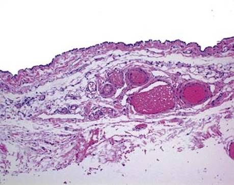
Fig 1 Pericardial cyst. Low-power view of pericardial cyst showing the mesothelial lining and cyst wall composed of loose connective tissue with blood vessels.

Fig 2 Pericardial cyst. Cyst lining; high-power view showing a single layer of mesothelial cells.