
Definition
• Benign neoplasm of peripheral nerve origin composed of Schwann cells, fibroblasts, and axons; occurring largely in the posterior mediastinum
Pathogenesis
• NF1 tumor suppressor gene on chromosome 17 has been implicated
• Patients with neurofibromatosis 1, an autosomal dominant disease, are heterozygous for NF1 mutation
• Loss of heterozygosity and “two-hit” hypothesis leads to inactivation of NF1 gene; new, sporadic mutations may also occur in patients without a family history of NF1
• Neurofibromin is a protein product of NF1, whose function is to inactivate RAS-GTP signal transduction pathways that promote cell proliferation and inhibit apoptosis
Clinical features
Epidemiology
• Accounts for about 5% of tumors of the mediastinum overall
• Comprising about 20% of mediastinal neurogenic tumors, it is the second most common tumor of this type after mediastinal schwannoma
• As with mediastinal schwannomas, lesions are commonly found in young adults; men and women are affected equally
• About 40% of mediastinal neurofibromas occur in patients with neurofibromatosis, who are usually seen at an earlier age
Presentation
• Patients are usually asymptomatic
• As with schwannomas in this region, larger lesions may result in compressive symptoms such as hoarseness, pain, or paresthesias
• Multiple solitary neurofibromas or plexiform neurofibromas are virtually diagnostic of neurofibromatosis
• MRI shows a homogenous spherical mass, which may less commonly show heterogeneity with cystic changes
Prognosis and treatment
• Primary treatment is surgical resection
• Recurrence of solitary neurofibroma after complete excision is rare
• Increased risk of malignant transformation of a neurofibroma in patients with neurofibromatosis
Pathology
Gross
• Tumors are extremely well-circumscribed and often lobulated to spherical. They are unencapsulated, in contrast to schwannomas. The cut surface is glistening and is softer than schwannomas
Histology
• Proliferation of all elements of the nerve, including Schwann cells, fibroblasts, and axons
• Variable amount of wavy, intersecting collagen fibrils, resembling “shredded carrots” between the dark-staining buckled nuclei of Schwann cells
• Often, a myxoid change with mucoid material separating the collagen
• Mast cells and lymphocytes are present in variable amounts
• Atypia and mitotic figures are rare
Immunopathology/special stains
• Immunohistochemical test against S100 protein shows only variable positivity in the neoplastic cells, demonstrating the heterogeneity of cell types within the tumor
• Immunohistochemical stain for neurofilament protein shows positive staining in the axons; this regular feature of neurofibroma can, however, also be observed in many schwannomas
Main differential diagnoses
• Schwannoma: usually encapsulated; Antoni A and Antoni B areas are present, and Verocay bodies may be found
• Malignant peripheral nerve sheath tumor: will demonstrate at least some mitotic activity, atypia, and often necrosis
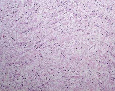
Fig 1 Mediastinal neurofibroma. Low-power view demonstrates a mixed population of cells in a “shredded carrot” loose collagen background.
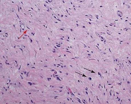
Fig 2 Mediastinal neurofibroma. Medium-power view shows a mixture of neoplastic Schwann cells with wavy nuclei (red arrow), fibroblasts, lymphocytes, and mast cells (black arrows).
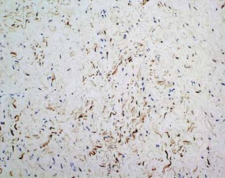
Fig 3 Mediastinal neurofibroma. Immunohistochemical stain for S100 protein shows positive staining in neoplastic Schwann cells only, with negative staining for fibroblasts and inflammatory cells.

Fig 4 Mediastinal neurofibroma. Immunohistochemical stain for neurofilament highlights entrapped axonal processes within the tumor; those cut in cross-section appear as round dots.

Fig 5 Mediastinal neurofibroma. Immunohistochemical stain for CD34 demonstrates not only blood vessels but also the extensive network of endoneurial fibroblasts, indicating the heterogenous nature of the cell population in this lesion: low power (A), high power (B).