
Definition
• A rare malignant neoplasm of peripheral nerve origin occurring largely in the posterior mediastinum
Pathogenesis
• Mutations have been detected in the NF1 and/or NF2 genes; these lead to tumorigenesis but are insufficient for malignant transformation
• Downregulation of tumor suppressor genes p53 and p16 occurs along with upregulation of epidermal growth factor receptor and platelet-derived growth factor receptor alpha
Clinical features
Epidemiology
• A rare tumor of the mediastinum
• Men and women are affected equally; most patients are in their third to fifth decades
• About half of all patients with MPNST have neurofibromatosis (NF)
• Conversely, approximately 5% of patients with NF go on to develop MPNSTs; these patients are usually seen as teens or young adults
• Tumors arise from simple or, more commonly, plexiform neurofibromas and rarely from schwannomas
Presentation
• In contrast to its benign counterparts, these tumors often have presenting symptoms of pain or nerve deficits
• The mass is usually large, greater than 5 cm, and may displace intrathoracic structures leading to hoarseness or respiratory distress
• Radiologically, a well-circumscribed mass may demonstrate local invasion of nearby structures
• Often, central necrosis and focal calcification are present
• Hematogenous metastasis to the lungs is common and more likely with NF patients
Prognosis and treatment
• Primary treatment is surgical resection; little benefit is derived from irradiation or chemotherapy
• Local recurrence after resection is common
• Overall prognosis is poor; 1-year survival is 33%
• Prognostic factors include tumor size, completeness of resection, and association with NF
Pathology
Gross
• The lesion causes enlargement of the nerve with the cut surface demonstrating a fairly well-circumscribed fleshy, tan appearance
• Central necrosis and/or hemorrhage is often present
• A pseudocapsule may be present
Histology
• Spindle cells are arranged in a fascicular pattern with large, wavy nuclei
• Frequent mitoses and sometimes bizarre cells
• Bony or cartilaginous metaplasia may be present
• Necrotic areas are surrounded by tumor cells in a palisading pattern
• Low-grade MPNSTs are less cellular, have a lesser degree of mitotic activity, and do not show as much necrosis
• About 5% of tumors have an epithelioid pattern consisting of plump, round cells with eosinophilic cytoplasm
Immunopathology/special stains
• Immunohistochemical test against S100 is focal in spindle cell MPNST but is diffusely positive in the epithelioid type
• p53 and CD99 may show positivity
Main differential diagnoses
• Schwannoma: Antoni A and Antoni B areas are present, and Verocay bodies may be found. Necrosis, atypia, and mitotic activity are not appreciable
• Neurofibroma: no atypia, necrosis, or mitoses
• Other sarcomas should be considered if tumor shows very weak or no S100 staining

Fig 1 Mediastinal malignant peripheral nerve sheath tumor. Disordered architecture of atypical neoplastic spindle cells admixed with fibroblasts and inflammatory cells in a collagenous background.
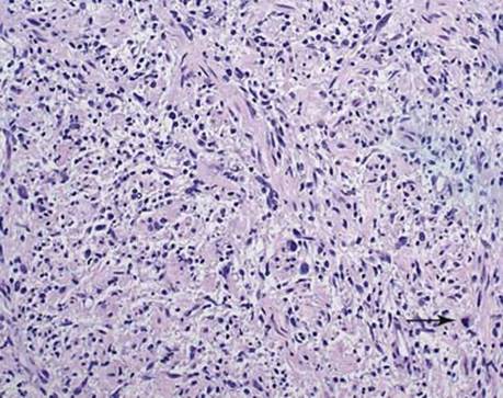
Fig 2 Mediastinal malignant peripheral nerve sheath tumor. Medium-power view shows haphazard arrangement of neoplastic cells. Note atypical mitotic figure (arrow).

Fig 3 Mediastinal malignant peripheral nerve sheath tumor. High-power view with bizarre, sometimes rhabdoid neoplastic cells (arrow).

Fig 4 Mediastinal malignant peripheral nerve sheath tumor. In this view, a large area of necrosis is evident next to viable tumor.

Fig 5 Mediastinal malignant peripheral nerve sheath tumor. Immunohistochemical stain for S100 protein highlights the enlarged, sometimes bizarre nuclei of malignant Schwann cells.