
Definition
• Rare neoplasm of neural crest chromaffin cell origin associated with the paraganglia of the sympathetic and parasympathetic nervous systems
Pathogenesis
• There is association with various hereditary syndromes such as multiple endocrine neoplasia type 2 (MEN2A and MEN2B), neurofibromatosis type 1, and von Hippel-Lindau disease
• Recently discovered germline mutations in familial paragangliomas include those involving succinate dehydrogenase (SDH) subunits
• SDH mutations account for more than 50% of familial cases and about 10% of sporadic cases
• Mutations of certain subunits have prognostic significance (e.g., presence of SDHD mutation is thought to confer a more malignant behavior)
• Anterior mediastinal lesions are thought to be associated with the aorticopulmonary paraganglia at the base of the heart, whereas those of the posterior mediastinum originate from the aorticosympathetic paraganglia at the costovertebral sulcus
Clinical features
Epidemiology
• Of all paragangliomas, the average age at presentation is 45 years
• Mediastinal lesions account for approximately 2% of all paragangliomas; conversely, paragangliomas account for <1% of mediastinal tumors
• Men and women are about equally affected
Presentation
• About half of patients are asymptomatic and are given a diagnosis after radiological chest imaging for other reasons
• Catecholamine secretion is rare in mediastinal paragangliomas but is more often associated with paravertebral (aorticosympathetic) tumors. Symptoms may include tachycardia, tremors, flushing, headache, and diaphoresis
• Nonsecreting tumors often reach large sizes and may be recognized because of mass effect
• Radiologically, CT and MRI are indicated; 123I-metaiodobenzylguanidine scintigraphy and/or PET-CT with 18-fluordeoxyglucose may be used to help stage the extent of disease
Prognosis and treatment
• Surgical resection offers a cure; chemotherapy and radiation therapy are not effective
• Prognosis after complete resection is good: 15-year survival rate is about 85%
• Distant metastasis may be seen in up to 10% of cases
Pathology
Gross
• Tumors are of variable size and show partial encapsulation. On sectioning, fleshy to firm areas are seen with some hemorrhage and/or fibrosis and a gray-brown cut surface
Histology
• Classically, the tumor shows a “zellballen” architecture of cells with nests surrounded by delicate, sometimes abundant, fibrovascular stroma
• The nuclei may be round and uniform or show pleomorphism with dense, sometimes vesicular chromatin
• Cytoplasm is granular and eosinophilic to amphophilic
• Mitoses and lymphovascular invasion may be seen but are not definitively associated with aggressive behavior or metastatic potential
• Melanin pigment and osseous metaplasia have been reported in the literature
Immunopathology/special stains
• The tumor cells may stain positively for neuron-specific enolase, chromogranin, synaptophysin, neurofilaments, and various peptides such as serotonin and somatostatin
• The surrounding sustentacular (supporting stromal) cells stain for S100 protein
• Keratin stains are almost always negative
Main differential diagnoses
• Neuroendocrine carcinoma: will have large cells, brisk mitotic activity, and positive keratin stains (e.g., CAM5.2)
• Metastatic carcinoma: may not have zellballen pattern; cytokeratins will be positive
• Melanoma: may not have zellballen pattern; melanoma markers (Melan-A, HMB45) will be positive
• Alveolar soft part sarcoma: cells are large with prominent nucleoli; MyoD1 shows cytoplasmic staining; PAS with diastase will demonstrate needle-like structures

Fig 1 Mediastinal paraganglioma. Low-power view shows an encapsulated lesion with dilated vessels.

Fig 2 Mediastinal paraganglioma. Medium power demonstrates nested zellballen architecture. Note the fine capillary network.
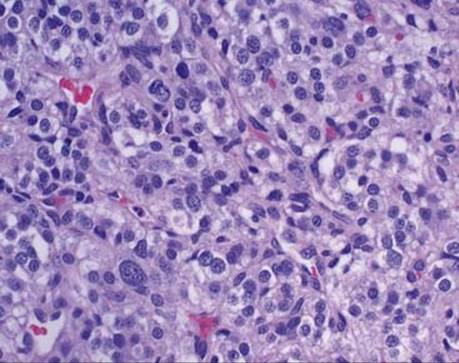
Fig 3 Mediastinal paraganglioma. High-power view shows nuclei of variable sizes with “salt and pepper” chromatin pattern and inconspicuous nucleoli. The cytoplasm is eosinophilic and granular. Some cells are dark and some are large, highlighting the presence of “endocrine atypia.”

Fig 4 Mediastinal paraganglioma. Immunostain for chromogranin demonstrates strong cytoplasmic staining.

Fig 5 Mediastinal paraganglioma. Immunostain for synaptophysin is positive in the cytoplasm and highlights the nested pattern of tumor cells.
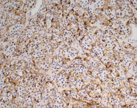
Fig 6 Mediastinal paraganglioma. Anti-S100 protein stains the supporting sustentacular cells around the nests of tumor.