
Definition
• T-cell lymphoma consisting of neoplastic large lymphoid cells that express anaplastic lymphoma kinase (ALK) protein and CD30 and are positive for the translocation involving the ALK gene (ALK negative ALCL is very rare and not discussed here)
Pathogenesis
• ALCL, ALK+ shows clonal rearrangement of the T-cell receptor genes. The ALK gene encodes a tyrosine kinase receptor, which is normally silent in lymphoid cells. In the presence of an abnormal fusion partner, most commonly nucleophosmin, a housekeeping gene [t(2,5)(p23;q35)], the formation of the heterodimer results in catalytic activation of the ALK domain and in the oncogenic properties of the ALK protein
Clinical features
Epidemiology
• ALCL, ALK+ accounts for approximately 3% of all adult non-Hodgkin lymphomas and 10% to 20% of the childhood lymphomas. ALCL, ALK+ is most frequent in the first three decades of life and has a male predominance
Presentation
• ALCL, ALK+ involves nodal and extranodal sites including skin, bone, soft tissues, lung, and liver. Mediastinal disease occurs less frequently than in classic Hodgkin lymphoma (cHL). Bone marrow involvement can be seen in up to 30% of cases, especially with aid of immunostains
• Majority of patients present with stage III-IV disease with peripheral and abdominal lymphadenopathy, often associated with extranodal infiltrates and bone marrow involvement. B-symptoms are also common
Prognosis and treatment
• Small-cell variant of ALCL has a more adverse outcome as it often presents with disseminated disease
• Overall 5-year survival of ALCL, ALK+ approaches 80%, while that of ALCL, ALK– tumors is around 48%. Relapse occurs in about 30% of cases but remains sensitive to chemotherapy
Pathology
Histology
• ALCL, ALK+ shows a broad morphological spectrum; variable number of cells with eccentric, horseshoe- or kidney-shaped nuclei, often referred to as hallmark cells. Hallmark cells are typically large but smaller forms may be seen. Some cells may show nuclear inclusions (invaginations of nuclear membrane), termed doughnut cells
• Multiple morphological patterns are recognized but have no clinical significance
• In the “common” pattern (60% of cases), tumor cells have abundant clear, basophilic, or eosinophilic cytoplasm. Wreathlike nuclear pattern may be seen resembling Reed-Sternberg cells. Tumor cells characteristically grow within sinuses, resembling metastasis.
• Other patterns include lymphohistiocytic pattern (10%), small-cell pattern (5%-10%), and the Hodgkin-like pattern (3%)
• More than one pattern may be seen in a single lymph node biopsy
• Immunophenotypically, tumors are positive for CD30 on the cell membrane and in the Golgi region. Large cells show strong immunoreactivity and cluster around blood vessels. The tumor cells show cytoplasmic and nuclear reactivity for ALK. In the small-cell variant, the ALK positivity is restricted to the nucleus. Vast majority of the cases are positive for epithelial membrane antigen (EMA) and one or more T-cell markers. CD3 is negative in more than 75% of cases.
Main differential diagnoses
• Hodgkin lymphoma: expression of CD15 and detection of weak PAX-5 expression on the neoplastic cells is critical in establishing the diagnosis of cHL. Polymerase chain reaction is often uninformative because the proportions of neoplastic cells may be below the limits of sensitivity in both cHL and ALCL
• Metastatic carcinoma: keratin stains often help resolve this differential
• Rhabdomyosarcoma and inflammatory fibroblastic tumors: rarely positive for ALK but are morphologically distinguishable from ALCL. In addition, these tumors tend to be negative for CD30 and EMA. A proportion of inflammatory myofibroblastic tumors additionally harbor increased numbers of plasma cells expressing the IgG4 subtype of IgG
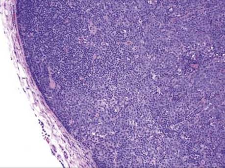
Fig 1 Anaplastic large-cell lymphoma of the mediastinum. Low-power view of the lymph node showing a sinusoidal infiltration by neoplastic cells.

Fig 2 Anaplastic large-cell lymphoma of the mediastinum. High-power view shows cells with irregular nuclear contours, abundant cytoplasm, and prominent nucleoli. Note the multiple abnormal mitotic figures in this field.
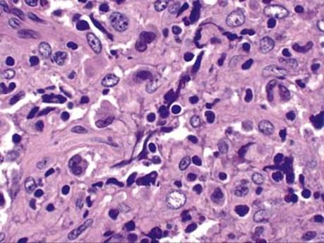
Fig 3 Anaplastic large-cell lymphoma of the mediastinum. High power from another case with large, pleomorphic cells, few of which show hyperchromatic horseshoe-shaped nuclei.
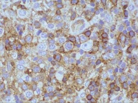
Fig 4 Anaplastic large-cell lymphoma of the mediastinum. High-power view of CD4 stain, showing strong immunoreactivity in the tumor cells supporting their T-cell origin.
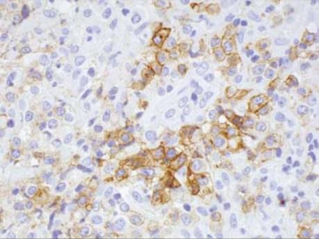
Fig 5 Anaplastic large-cell lymphoma of the mediastinum. The tumor cells show membrane and cytoplasmic staining for CD30.

Fig 6 Anaplastic large-cell lymphoma of the mediastinum. The tumor cells also show strong immunoreactivity for leukocyte common antigen.
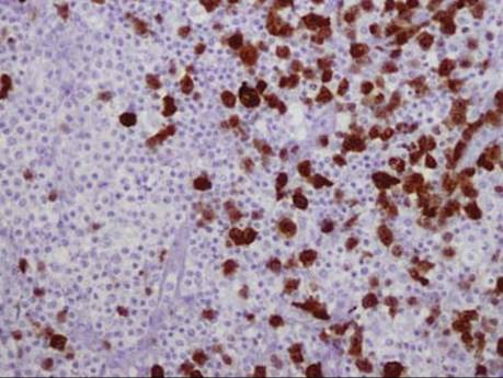
Fig 7 Anaplastic large-cell lymphoma of the mediastinum. Strong staining (both nuclear and cytoplasmic) for ALK protein is noted in the majority of the neoplastic cells.