
Definition
• Rare neoplastic condition characterized by tumor-forming masses composed of immature myeloid cells, at an extramedullary site
• Synonyms and older terms include extramedullary acute myeloid leukemia, extramedullary myeloid tumor, granulocytic sarcoma or chloroma
Pathogenesis
• MS may develop de novo or concurrently with acute myeloid leukemia (AML), myeloproliferative neoplasm (MPN), or myelodysplastic syndrome (MDS)
• MS may be the first manifestation of AML (even precede it by months or years) or may represent the initial manifestation of relapse in a previously treated AML in remission
• At the cytogenetic level, the most common reported association is with MLL rearrangement and translocation t(8;21). In a single large study that systematically evaluated MS by fluorescence in situ hybridization, several aberrations were detected, including monosomy 7, trisomy 8, MLL splitting, inv(16), trisomy 4, monosomy 16, 16q–, 5q–, 20q–, and trisomy 11. About 16% of cases carry nucleophosmin (NPM) mutations, as shown by aberrant cytoplasmic NPM expression; these cases usually show myelomonocytic or monocytic morphological features and a normal karyotype
Clinical features
Epidemiology
• A rare neoplasm, with very few case series; epidemiological details are not well characterized
Presentation
• The most frequent sites are bone, lymph node, or skin, although any part of the body may be affected
• Patients may present with nonspecific systemic symptoms (such as fever) or with features related to the tumor masses (such as lymphadenopathy and subcutaneous nodules)
Prognosis and treatment
• Reported 5-year survival rate has usually been below 25%. There are no significant differences in terms of survivorship between the groups with de novo tumor and the one with concomitant or previous hematological disorders (AML, MPN, or MDS), although smaller studies have suggested better outcomes in those with de novo disease
• Patients who undergo allogeneic or autologous bone marrow transplantation seem to have a higher probability of prolonged survival or cure. Response to first-line therapy affects outcome
• Clinical behavior and response to therapy was not influenced by age, sex, anatomical site, histological features, immunophenotype, or cytogenetic findings
Pathology
Histology
• Architecturally, MS forms a tumor mass with sheets of neoplastic cells. In lymph nodes, the neoplastic cells may diffusely infiltrate sinuses or the paracortex, entrapping some residual follicles. At extranodal sites, the tumors may show a diffuse or Indian-file growth pattern, depending on the degree of stromal reaction evoked
• The neoplastic cells are mostly myeloblasts, with or without features of promyelocytic or neutrophilic maturation. A significant proportion of cases display monoblastic, monocytic, or myelomonocytic differentiation. Trilineage hematopoiesis or a predominance of erythroid precursors or megakaryoblasts is rare and may point to prior chronic MPN or MDS
• The recent World Health Organization classification has proposed the subdivision of MS into several types
Immunopathology/special stains
• CD68-KP1 is the most commonly expressed marker followed by myeloperoxidase (MPO) and CD117
• Other markers that are positive include CD99, CD68/PG-M1, lysozyme, CD34, terminal deoxynucleotidyl transferase (TdT), CD56, CD61/linker of activated T lymphocyte/factor VIII–related antigen, CD30, glycophorin A, and CD4
• Foci of plasmacytoid dendritic cell differentiation (CD123+) may be observed in cases carrying inv(16)
• The proliferation index (Ki-67/MIB1 score) is high, ranging from 50% to 95%
Main differential diagnoses
• On morphological grounds alone, the lesion may be misdiagnosed as lymphoblastic lymphoma, Burkitt lymphoma, diffuse large B-cell lymphoma, small-cell carcinoma, or other small round blue cell tumors (such as rhabdomyosarcoma or Ewing sarcoma). Immunohistochemical workup helps differentiate these entities
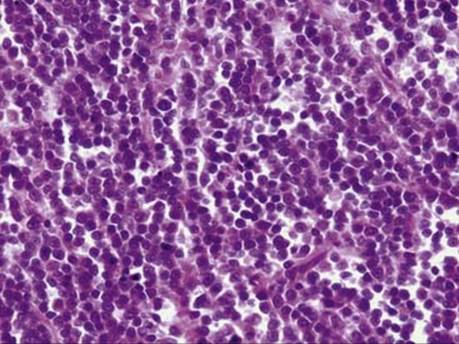
Fig 1 Myeloid sarcoma. This typical MS shows sheets of round cells with blastic morphology.
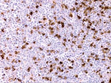
Fig 2 Myeloid sarcoma. The neoplastic cells show strong positivity for MPO.

Fig 3 Myeloid sarcoma. This patient presented with pericardial effusion, and a pericardial mass was resected. The tumor was composed of poorly differentiated pleomorphic and round cells (A) that were strongly positive for MPO (B) and also showed monocytoid differentiation as evidenced by lysozyme positivity (C).