
Definition
• Also known as angiofollicular lymph node hyperplasia and giant lymph node hyperplasia; is a nonclonal lymphoproliferative disorder that can affect single lymph nodes or have a multicentric presentation
Pathogenesis
• IL-6 stimulation has been implicated in the pathogenesis; it induces a CD-like condition in mice. In patients with HIV, human herpesvirus (HHV)-8 induces a viral IL-6 homolog that is implicated in the pathogenesis through induction of angiogenesis and hematopoiesis with consequent systemic manifestations and is termed multicentric Castleman disease (MCD). The role of HHV-8 in non-HIV patients is more controversial. The IL-6 upregulation is also implicated in the pathogenesis of pulmonary arterial hypertension seen in some patients with CD
Clinical features
Epidemiology
• The first case, described by Dr. Benjamin Castleman in 1954, was a patient with solitary hyperplastic mediastinal nodes with regressive germinal centers; characterized as unicentric CD
• Subsequent studies noted multicentric lymphadenopathies characterized by constitutional symptoms with histological features similar to unicentric CD
• Subsequently, HHV-8 was identified in MCD, particularly in HIV-positive patients
Presentation
• Median age of presentation is in the fourth decade; equal incidence in men and women
• Thoracic involvement by CD usually seen as hilar/mediastinal lymphadenopathy; rarely, interstitial infiltrates within the lung parenchyma
• Following types have been described:
• Hyaline–vascular subtype seen usually as a unicentric mass lesion with lack of systemic signs and symptoms. Associated lesions include follicular dendritic neoplasms, vascular/stromal neoplasms
• The plasma cell variant is more often multicentric and may represent a component of polyneuropathy, organomegaly, endocrinopathy, monoclonal gammopathy, skin changes (POEMS) syndrome
• HHV-8 associated MCD is known to occur in immunosuppressed and HIV-positive patients. HHV-8–associated multicentric CD has a higher risk of progression to HHV-8 positive plasmablastic lymphoma. Kaposi sarcoma (KS) also frequently occurs in this subgroup
• The multicentric type, not otherwise specified, and the plasma cell variant may be associated with POEMS syndrome and an increased risk of lymphomas in the former
• Generalized lymphadenopathy, constitutional symptoms, and hematological and immunologic abnormalities are more commonly associated with the plasma cell, HHV-8–associated, and multicentric variants
Prognosis and treatment
• HHV-8–associated MCD has a poor prognosis; survival, only a few months. May be an increased risk of other lymphomas in any patient with CD, regardless of HIV status
• Unicentric CD can be surgically excised or irradiated
• Multicentric CD is treated with chemotherapy, steroids, or rituximab
• Tocilizumab, a humanized anti-interleukin-6 receptor (IL-6R) monoclonal antibody, has been demonstrated in a few studies to ameliorate symptoms. In HHV-8 MCD, treatment of the associated HIV with highly active antiretroviral therapy also results in substantial clinical improvement
Pathology
Histology
• At least three distinct pathological types of Castleman’s disease:
• Hyaline vascular type (hvCD): lymph node follicles have widened mantle zones composed of concentric rings of small lymphocytes (onion-skin pattern). Often, more than one regressed follicle may be seen within a common expanded mantle cuff (“twinning”). The germinal centers are atrophic or atretic with penetrating hyalinized small vessels and prominent follicular dendritic cells. Occasional cases may show associated prominent angiomyoid proliferations or dysplastic follicular dendritic cell proliferations within the same node
• Plasma cell variant type: retained lymph node architecture, hyperplastic germinal centers with sheets of plasma cells in the interfollicular and paracortical region, increased vascularity, and expanded mantle zones
• Plasmablastic variant: seen in the context of HHV-8 MCD; shows multiple varying sized reactive or regressed germinal centers with singly scattered plasmablasts within the mantle zones; varying amount of interfollicular plasmacytosis. In a significant proportion of cases, there may be associated KS either within the same node or adjacent nodes
Immunopathology/special stains
• hvCD: CD23 and CD21 will mark the tight follicular dendritic cell meshworks; EGFR often marks dysplastic dendritic cells, especially when follicular architecture is obliterated, making identification of follicular dendritic cells difficult
• Plasma cell variant: rarely monoclonal plasma cell proliferations can be present in this variant, and kappa and lambda immunostains help identify this component
• HHV-8 MCD: the plasmablasts in the mantle zones express monoclonal IgM/lambda immunostain besides nuclear HHV-8. In addition, there are polyclonal interfollicular plasma cells. HHV-8 stain is often useful for identifying areas of KS not obvious on H&E staining
Main differential diagnoses
• Rheumatic and viral induced lymphoid hyperplasia
• Dysplastic follicular dendritic cells can mimic Hodgkin lymphoma, which is negative for CD21, CD23, and EGFR stains
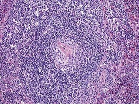
Fig 1 Mediastinal Castleman disease. Low power; H&E image of a residual lymphoid follicle showing a hyalinized blood vessel penetrating its center (the “lollipop lesion”).
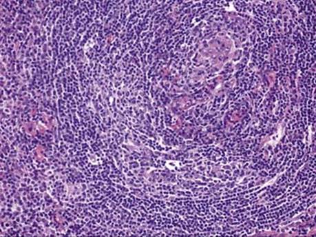
Fig 2 Mediastinal Castleman disease. H&E image of a lymphoid follicle with prominent mantle zone composed of concentric rings of small lymphocytes (onion-skin pattern).
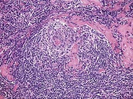
Fig 3 Mediastinal Castleman disease. H&E image showing a regressed and devascularized germinal center.