
Definition
• Benign malformation of dilated lymphatic channels
Clinical features
Epidemiology
• Comprises up to 5% of mediastinal masses
• More common in pediatric patients
• In contrast to cystic lymphangiomas (cystic hygromas) of the neck, which be associated with Turner syndrome, there is no known association with mediastinal lymphangiomas
• Can be an isolated lesion or can occur in patients with lymphangiomatosis
Presentation
• In pediatric patients, it is usually seen as an anterosuperior mediastinal mass with a cervical component. Adults usually lack the cervical component
• Most cases are asymptomatic and found incidentally on imaging studies
• When present, symptoms may include dyspnea, coughing, hemoptysis, chest pain, dysphagia, upper extremity swelling
Prognosis and treatment
• Prognosis is excellent
• Treatment is complete surgical excision
Pathology
Histology
• Irregular spaces lined by flat lymphatic endothelial cells
• Lumina may be filled with proteinaceous fluid, lymphocytes, or erythrocytes
• Loose connective tissue stroma often with lymphocytic infiltrate
• Mast cells and hemosiderin deposits in the stroma are common
• Scattered bundles of smooth muscle in the walls of larger channels
• May incorporate surrounding adipose tissue, blood vessels, and lymph nodes
Immunopathology/special stains
• D2-40: membranous stain highlights lymphatic endothelial cells
• Variable expression of CD31, CD34, and FVIII in endothelial cells
Main differential diagnoses
• Hemangioma: endothelial cells are CD34 positive (membranous) and D2-40 negative
• Bronchogenic cyst: lined by ciliated columnar epithelium. May have islands of cartilage, smooth muscle fascicles, and scattered bronchial glands. Ulceration of epithelial lining can occur, making it difficult to distinguish from other lesions
• Pericardial cyst: lined by flat to cuboidal mesothelial cells; strongly keratin positive

Fig 1 Mediastinal lymphangioma. Dilated lymphatic channels in connective tissue stroma.

Fig 2 Mediastinal lymphangioma. Smooth muscle is present in the walls of these larger lymphatic channels, which are filled with proteinaceous material. Also present are incorporated small blood vessels and adipose tissue (low power).

Fig 3 Mediastinal lymphangioma. Medium-power view of Fig 2 showing smooth muscle in the walls of these larger lymphatic channels.
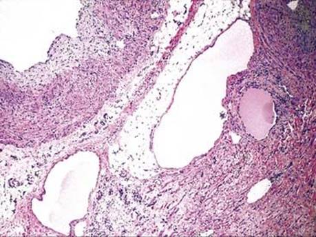
Fig 4 Mediastinal lymphangioma. Dilated lymphatic channels and a lymphocytic infiltrate within the connective tissue stroma. The large channel in the upper left has a thick smooth muscle wall.
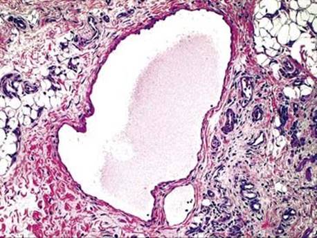
Fig 5 Mediastinal lymphangioma. Dilated lymphatic channels, connective tissue stroma with scattered lymphocytes, incorporated blood vessels, and adipose tissue are seen here at high power.

Fig 6 Mediastinal lymphangioma. Dilated lymphatic channel with adjacent lymphocyte aggregate.

Fig 7 Mediastinal lymphangioma. Immunohistochemistry with D2-40 highlights the lymphatic endothelial cells.

Fig 8 Mediastinal lymphangioma. High power showing positive membranous staining of lymphatic endothelial cells with D2-40.