
Definition
• Malignant mesenchymal neoplasm composed at least in part of immature or mature adipocytes
Clinical features
Epidemiology
• Rare; accounts for <1% of mediastinal tumors and 9% of primary sarcomas in the mediastinum
• Occurs in adults ages 20 to 70 years (average, fifth decade)
Presentation
• Often remains asymptomatic until it reaches a very large size
• Most common symptoms include dyspnea, tachypnea, wheezing, coughing
• Less commonly presents with acute pericarditis, cardiac tamponade, syncope, angina, pleural effusion, metastases, superior vena cava obstruction
• Usually located in the posterior mediastinum; rare cases reported in the anterior mediastinum
Prognosis and treatment
• Ideally, complete surgical excision is performed
• If unable to completely resect due to location or infiltration of adjacent structures, adjuvant chemotherapy or radiation therapy may be used
• Tumor behavior correlates with histological subtype: dedifferentiated and poorly differentiated tumors are more aggressive and well-differentiated and myxoid tumors have a better prognosis
• Myxoid tumors are highly sensitive to chemotherapy and radiation therapy
Pathology
Gross
• Circumscribed, multilobulated tumor
• Cut surface is yellow to tan-gray depending on histological subtype and proportion of elements. Myxoid areas will have a gelatinous appearance
• Large tumors may have areas of fat necrosis
Histology
• Well-differentiated
• Composed of mature adipocytes of varying sizes
• Focal adipocyte nuclear atypia and hyperchromasia
• Occasional atypical stromal cells with hyperchromatic nuclei and/or multinucleation; most prominent in the fibrous septa
• Monovacuolated or multivacuolated lipoblasts may be present but are not required for the diagnosis. Multivacuolated lipoblasts have a scalloped nucleus, indented by the vacuoles
• Heterologous differentiation may occur with formation of bone, smooth or striated muscle
• Four main subtypes:
– Adipocytic: lipoma-like
– Sclerosing: extensive collagenous stroma
– Inflammatory: chronic inflammatory infiltrate
– Spindle cell: bland spindle cells with fibrous or myxoid stroma
• Myxoid
• Nodular pattern with increased cellularity at the periphery of the nodules
• Myxoid stroma with delicate branching “chicken wire” vasculature
• Pools of extracellular mucin create a microcystic or “pulmonary edema” pattern
• Mixture of round to oval mesenchymal cells and signet-ring lipoblasts
• Dedifferentiated
• Varying amounts of dedifferentiation, often with an abrupt transition from well to poorly differentiated areas
• Dedifferentiated areas usually resemble pleomorphic sarcoma or myxofibrosarcoma
• Low-grade dedifferentiation consists of fascicles of bland spindle cells without a lipogenic component
• Heterologous differentiation to myosarcoma, osteosarcoma, chondrosarcoma, or angiosarcoma may occur
• Meningothelial-like whorling pattern may be present
• Pleomorphic
• Pleomorphic spindle cells in fascicles
• Varying amounts of pleomorphic lipoblasts
• Eosinophilic hyaline droplets or globules are often present intracellularly and extracellularly
• Epithelioid variant is composed of sheets of tumor cells with well-defined cell borders; eosinophilic cytoplasm and prominent nucleoli separated by fibrous septae containing capillaries
• Mixed-type liposarcoma
• Shows a combination of features of myxoid liposarcoma with well-differentiated, dedifferentiated, or pleomorphic liposarcoma
Immunopathology/special stains
• Well-differentiated: MDM2+, CDK4+
• Myxoid: diffusely S100+
• Dedifferentiated: MDM2+, CDK4+
• Poorly differentiated: vimentin+; 50% are S-100+
Main differential diagnosis
• Spindle cell/pleomorphic lipoma: ropey collagen, “floret-like” multinucleated giant cells, strongly CD34+
• Lipoblastoma: usually seen younger than age 5; prominent lobules separated by connective tissue; myxoid stroma and plexiform vascular pattern mimic myxoid liposarcoma, but it lacks giant cells and pleomorphic nuclei
• Myxofibrosarcoma: prominent curvilinear thin-walled blood vessels; vacuolated pseudolipoblasts

Fig 1 Mediastinal liposarcoma. Well-differentiated lipoma-like liposarcoma with mostly mature-appearing adipocytes. Interspersed atypical nuclei are easily seen even at this low power.

Fig 2 Mediastinal liposarcoma. Lipoblast with multiple vacuoles.

Fig 3 Mediastinal liposarcoma. Atypical stromal cells in a well-differentiated liposarcoma.
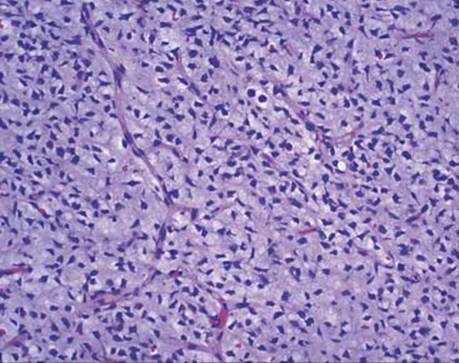
Fig 4 Mediastinal liposarcoma. High-power view of “chicken wire” vessels of a myxoid liposarcoma.
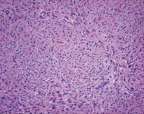
Fig 5 Mediastinal liposarcoma. Dedifferentiated liposarcoma composed of fascicles of spindled cells.

Fig 6 Mediastinal liposarcoma. Transition between well-differentiated and dedifferentiated liposarcoma.