
Gross anatomy
Weight
• In females, 250 g to 300 g
• In males, 300 g to 350 g
General orientation
• Ventricles are anterior
• Great vessels are anterior to the atria
• Posterior surface is flat
Coronary arteries
• Left main coronary artery: arises from the aorta and divides into the left anterior descending (LAD) and left circumflex (LCX)
• LAD: travels down the anterior surface of the heart in the interventricular groove
• LCX: travels in the groove between the left atrium and ventricle
• Right main coronary artery: runs between the base of the pulmonary artery and right auricle and continues in the right atrioventricular groove
Right atrium
• Receives blood from the superior and inferior vena cavae and the coronary sinus
• Right atrial appendage (auricle) is triangular with a broad base
• Crista terminalis: muscular ridge separating the smooth-walled portion of the atria from the prominent ridges of the pectinate muscles
Right ventricle
• Thicker trabeculae in a more disorganized pattern than the left ventricle
• Thick muscular wall between inflow and outflow tracts
• Normal wall thickness: 0.3 cm to 0.5 cm
Left atrium
• Left atrial appendage is wormlike with a narrow base
• Smooth endocardial surface
• Receives oxygenated blood from the four pulmonary veins
Left ventricle
• More delicate trabeculae oriented from the base toward the apex
• Thin connective tissue separating the inflow and outflow tracts
• Normal wall thickness: 1.3 cm to 1.5 cm
Valves
• Semilunar valves
• Aortic valve: normal circumference: females, 5.7 cm to 7.9 cm; males 6 cm to 8.5 cm
• Pulmonary valve: normal circumference: females, 5.7 cm to 7.4 cm; males, 9.2 cm to 9.9 cm
• Have three semilunar cusps
• Atrioventricular valves
• Mitral (bicuspid): normal circumference: females, 8.2 cm to 9.1 cm; males, 9.2 cm to 9.9 cm
• Tricuspid: normal circumference: females, 10 cm to 11.1 cm; males, 9.2 cm to 9.9 cm
• Anchored to the ventricular wall by chordae tendinae attached to papillary muscles
Histology
Endocardium
• Single layer of endothelial cells
Myocardium
• Bundles of cardiac myocytes separated by fibrous bands
• Cardiac myocytes
• Single centrally located nuclei
• Cross striations
• Branching
• In infants and children, the myocytes are smaller and the cytoplasm has more glycogen than in adults
• Intercalated discs: thin dark pink line perpendicular to the myocyte fibers marking the junction between adjacent myocytes
Epicardium (visceral pericardium)
• Single layer of mesothelial cells with underlying basement membrane and varying amount of fat
Valves composed of three layers
• Fibrosa: dense collagen
• Spongiosa: loose collagen fibers, fibroblasts, proteoglycans
• Ventricularis: elastic fibers
Embryology
• Angiogenic clusters in the splanchnic mesoderm form two endocardial tubes
• Day 19: fusion of two tubes to form a single cardiac tube
• The heart tube has five dilations, which are from superior to inferior the truncus arteriosus, bulbus cordis, primitive ventricle, primitive atria, sinus venosus
• Day 22 (range, 19 to 24): heart begins beating
• Day 23: looping of the heart
• Bulbus cordis moves inferiorly, anteriorly, and to the right
• Primitive ventricle moves left
• Primitive atria and sinus venosus move superior and posterior
• Truncus arteriosus separates into the aorta and pulmonary artery by growth and fusion of spiral truncoconal ridges
• Fourth week: ventral and dorsal endocardial cushions extend and fuse, separating the heart into atria and ventricles
• Fifth week: septum primum grows down from the top of the atria and merges with the endocardial cushions to form the interatrial septum
• Foramen primum: space between leading edge of septum primum and endocardial cushions
• Foramen secundum: foramen that forms superior to the foramen primum via apoptosis as the septum primum fuses with the endocardial cushions
• Septum secundum forms to the right of the septum primum. It contains an opening, the foramen ovale, to allow for blood flow between the atria
• Interventricular septum
• Muscular interventricular septum forms as a wall that grows up from the apex of the common ventricle toward the endocardial cushions and stops inferior to the atrioventricular septum, creating an interventricular foramen
• Membranous interventricular septum forms from growth of the bulbar ridges and endocardial cushions and fusion of these with the muscular septum

Fig 1 Normal heart. This gross image shows the right main (RCA) and left main (LCA) coronary arteries coming off the ascending aorta.

Fig 2 Normal heart. The LAD artery is seen traveling down the anterior aspect of the heart in the interventricular groove. The LCX artery branches off the left main coronary artery and travels under the left auricle in the left atrioventricular groove.

Fig 3 Normal heart. The posterior descending artery (PDA) branches from the right coronary artery (RCA).

Fig 4 Normal heart. Low-power microscopic image of normal cardiac myocytes cut longitudinally.

Fig 5 Normal heart. High-power microscopic image of normal cardiac myocytes. Note presence of lipofuscin in the cytoplasm, adjacent to the nucleus, in this heart from a 51-year-old female.
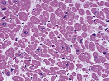
Fig 6 Normal heart. Cardiac myocytes have a single centrally located nucleus and eosinophilic cytoplasm seen here on cross-section.

Fig 7 Normal heart. Increased amount of glycogen in the myocytes of an infant is seen here. Because the fibers are small, the section appears hypercellular.