
Definition
• Cardiomyopathy characterized by ventricular or four-chamber dilation and systolic dysfunction with impaired contraction of one or both ventricles
• Significant coronary artery disease, hypertensive heart disease, and valvular disease are absent
Pathogenesis
• About 50% of cases are idiopathic, 25% to 35% are familial, and the remainder are due to chronic alcohol abuse, heavy metal toxicity, and pregnancy
• In idiopathic cases, infectious causes of myocarditis or autoimmune mechanisms are suspected
• In familial cases, multiple genetic mutations are involved, including genes coding sarcomeric, cytoskeletal, and nuclear proteins, as well as proteins that regulate calcium metabolism
• Alcohol and its metabolites are toxic to myocytes, but no features distinguish alcoholic cardiomyopathy from other dilated cardiomyopathies
• Occurs late in pregnancy or weeks to months postpartum and may be due to pregnancy-associated hypertension and volume overload
Clinical features
Epidemiology
• Estimated total prevalence: 1 in 2500
• Typically seen in patients between 20 and 60 years old but can also occur in children
Presentation
• Symptoms of heart failure: dyspnea on exertion, peripheral edema, orthopnea, paroxysmal nocturnal dyspnea
• May present with a slow progression to heart failure but can rapidly progress to a decompensated state
• Can be incidentally detected with asymptomatic cardiomegaly, conduction disturbances and arrhythmias, thromboembolic complications, or sudden death
Prognosis and treatment
• About 50% of patients die within 2 years; only 25% survive longer than 5 years
• Death due to progressive heart failure, arrhythmia, or emboli
• Idiopathic dilated cardiomyopathy is the most common indication for heart transplantation
Pathology
Gross
• Ventricular or four-chamber dilation
• Globular-shaped heart from dilation
• Increased heart weight from hypertrophy
• Normal or reduced left ventricular free-wall thickness
• Mural thrombi in one or more chambers
• Prominent reticulation of left ventricle trabeculations
• Fibrous thickening of endocardium of the septum
Histology
• Myocyte hypertrophy with enlarged, irregular, hyperchromatic nuclei
• Mild interstitial and perivascular fibrosis
• Hypertrophic myocytes with increased vacuolization
• Focal interstitial lymphocytes and macrophages without myocyte damage
Immunopathology/special stains
• Not contributory
Main differential diagnoses
• Endomyocardial biopsy to distinguish from lymphocytic myocarditis, sarcoidosis, and hemochromatosis
• Detailed clinical history to distinguish from vascular disorders, endocrine causes, nutritional deficiencies, and recreational drug abuse
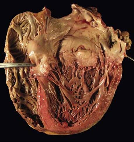
Fig 1 Dilated cardiomyopathy. Gross photograph of globular-shaped heart with four-chamber dilation seen in dilated cardiomyopathy.

Fig 2 Dilated cardiomyopathy. Hypertrophic myocytes with enlarged, irregular, and hyperchromatic nuclei seen in dilated cardiomyopathy: low (A) and high (B) powers.
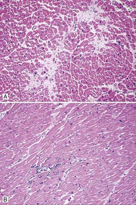
Fig 3 Dilated cardiomyopathy. Focal interstitial fibrosis (A) and lymphocytes (B) seen in dilated cardiomyopathy.
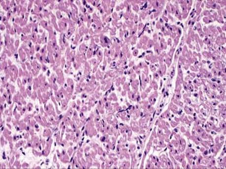
Fig 4 Dilated cardiomyopathy. This 2-year-old underwent transplantation for dilated cardiomyopathy. Note that although the fibers are small because of the young age, the nuclei are enlarged and hyperchromatic.