
Definition
• Cardiomyopathy characterized by hypertrophy of the left ventricle and abnormal diastolic filling
• Hypertensive disease, valvular disease, and systemic or infiltrative disorders are absent
Pathogenesis
• Eleven mutations that encode sarcomere proteins have been identified
• Mutations in cardiac myosin-binding protein C are the most common
• Mutations in cardiac beta-myosin heavy chain occur in approximately one third of cases
• Mutations in troponin I, troponin T, and alpha-tropomyosin are less common
• How these mutations effect contractility is not well understood
Clinical features
Epidemiology
• Prevalence: 1 in 500
• Most common genetic heart disorder
• Can be seen at any age, including infancy, but is more common in adolescence
Presentation
• Impaired diastolic filling and hypertrophic left ventricle resulting in reduced chamber size, poor compliance, and reduced stroke volume leading to limited cardiac output
• Outflow tract obstruction if there is contact between the septum and anterior mitral leaflet
• Majority of patients have minor or no symptoms
• Symptoms include exertional dyspnea, chest pain, palpitations, lightheadedness, syncope, and sudden death
• Common cause of sudden death in young athletes
Prognosis and treatment
• Mortality is due to sudden cardiac death, heart failure, and stroke
• Treatment is medical management with beta blockers or verapamil to slow the heart rate and increase diastolic filling
• Ventricular septal myotomy–myomectomy to remove septum beneath aortic valve in patients with severe outflow obstruction or in patients in whom medical management has failed
Pathology
Gross
• Marked asymmetric thickening of the intraventricular septum; rare cases with concentric hypertrophy
• Whorled appearance of septal wall musculature
• Normal or reduced left ventricle chamber volume
• Right ventricular wall thickening, particularly in the anterior wall of the outflow tract
Histology
• Marked myocyte hypertrophy with pleomorphic and hyperchromatic nucleoli
• Myocyte disarray
• Interstitial and perivascular fibrosis
• Endocardial fibrosis
• Intramyocardial arteries with smooth muscle proliferation and reduced lumens
Immunopathology/special stains
• Trichrome stain highlights interstitial and endocardial fibrosis
Main differential diagnoses
• Hypertensive heart disease
• Amyloidosis and other infiltrative disorders
• Valvular or congenital subvalvular aortic stenosis
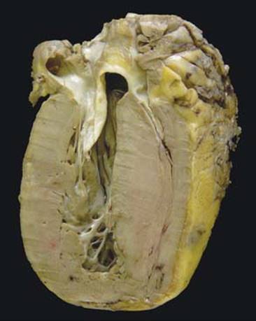
Fig 1 Hypertrophic cardiomyopathy. Gross photograph of hypertrophic cardiomyopathy with asymmetrically thickened left ventricle.

Fig 2 Hypertrophic cardiomyopathy. Hypertrophic myocytes with pleomorphic hyperchromatic nucleoli seen in hypertrophic cardiomyopathy.
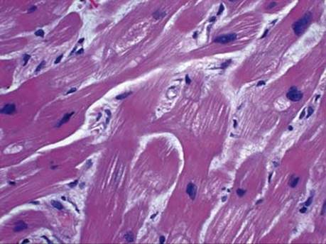
Fig 3 Hypertrophic cardiomyopathy. Myocyte disarray seen in hypertrophic cardiomyopathy.

Fig 4 Hypertrophic cardiomyopathy. Interstitial and perivascular fibrosis seen in hypertrophic cardiomyopathy.