
Definition
• Death of cardiac myocytes resulting from ischemia
Pathogenesis
• About 90% of cases have associated coronary vessel atherosclerosis (see chapter on Atherosclerosis for detailed discussion of its pathogenesis)
• Most often, cases of MI involve complications of the atherosclerotic plaque, such as acute rupture, hemorrhage within the plaque, or ulceration
• Subsequent platelet aggregation and activation at the site of plaque change results in thrombus formation that, within minutes, leads to complete occlusion of the vessel
• Most often there is transmural infarction; however, rapid lysis of the thrombus may result in only subendocardial injury
• Downstream ischemia of cardiac myocytes occurs. Because they are incapable of anaerobic glycolysis, they require a constant supply of glucose and oxygen from the bloodstream
• Lack of glucose and oxygen leads to accumulation of toxic products such as lactic acid; increased demand, as with tachycardia, or a change in hemodynamics, as with hypotension, increases the risk of an infarct
• Prolonged ischemia (greater than 30 minutes) leads to irreversible damage, with necrosis and cell death. Thus, permanent myocyte damage can be averted if measures are taken within this time frame (e.g., aspirin or thrombolytic administration)
• Ischemia greater than 3 hours may lead to necrosis of a significant area of myocardium, placing the patient in danger of fatal arrhythmias and late complications (see further on)
• Other causes of MI not related to atherosclerosis include vasospasm and paradoxical emboli
Clinical features
Epidemiology
• MI is the leading cause of death in the western world
• Over 1.5 million patients have MI every year; 500,000 of them die, often before reaching the hospital
• Risk factors include male gender, age (only 10% of MIs occur in those younger than 65 years), postmenopausal state (premenopausal women have hormone protection against atherosclerosis), and other risk factors related to atherosclerosis
Presentation
• Patients classically present with crushing substernal chest pain, diaphoresis, weak pulses, dyspnea, and referred pain to the jaw and/or the right arm
• Patients with diabetes mellitus and the elderly may have “silent” infarcts due to neuropathy
• Diagnosis is established using a combination of a good history and physical, electrocardiographic (ECG) findings, and laboratory evidence of myocardial damage
• Troponins T and I have high sensitivity and specificity and are considered the laboratory gold standard for diagnosis of acute MI; levels rise about 2 hours after injury, peak at 24 hours, and remain elevated for 7 to 10 days
• Cardiac-specific creatine kinase (CK-MB) levels also rise about 2 hours after injury and peak at 24 hours. However, levels normalize within 72 hours. Thus, CK-MB is most helpful in assessing reinfarctions, where troponins would still be elevated
Prognosis and treatment
• The outcome of an episode of MI is related to the size of the infarct, the promptness of treatment administration, and factors related to the patient’s cardiac status (e.g., previous MI or cardiac hypertrophy)
• About half of patients die before reaching the hospital; an additional 10% die in the hospital, often because of related complications of the MI
• Large infarcts lead to left ventricular dysfunction, some may manifest as cardiogenic shock, which carries a 70% mortality rate
• Many patients also have arrhythmias, a consequence of myocyte irritability secondary to cell injury
• Other complications include myocardial rupture (either of free wall, papillary muscle, or interventricular septum, all of which are due to muscle weakening after necrosis and inflammation, occurring 3 to 7 days after MI), pericarditis, ventricular aneurysm, or mural thrombi
• Treatment involves prompt intervention with aspirin, nitroglycerin, oxygen, and morphine. Reperfusion with tissue plasminogen activator (t-PA), angioplasty, or emergent coronary artery bypass grafting (CABG) may restore muscle that is injured but not yet dead
Pathology
Gross and microscopic morphology
• Changes to ischemic myocardium are often characterized based on time from vessel occlusion
• Before 4 hours, gross changes are not noticeable; microscopically, waviness of fibers may be seen
• By 24 hours, there is dark mottling of the myocardium grossly, along with neutrophilic infiltrate, edema, contraction band necrosis, and hemorrhage
• Within the first week, yellow-tan discoloration and parenchymal softening may be seen; microscopically, active phagocytosis of dead myocytes occurs
• By 2 weeks, a gray-white scar develops, often with depressed infarct borders; collagen and granulation tissue is deposited
• In lesions older than 2 months, a gray-white, stellate scar can be seen that histologically corresponds to deposition of dense collagenous tissue replacing the area of infarct; few residual myocytes remain
Immunopathology/special stains
• Immunohistochemical stain for C4d stains the cytoplasm of necrotic cardiac myocytes and was found to be positive even before the infiltration of inflammatory cells, allowing detection of early MI
• Triphenyltetrazolium chloride is a histochemical stain that utilizes the compound to indicate cell death. It is a white compound that, in the presence of dehydrogenases (e.g., lactate dehydrogenase [LDH]) turns red. In necrotic tissues where LDH is low, the area of infarction will remain yellow-white while the surrounding tissue will impart a dark red color. The stain is helpful in discerning an infarct that is at least 2 hours old
Main differential diagnosis
• Myocarditis: has prominent, usually lymphocytic, infiltrate; atherosclerosis and/or coronary artery disease often are not present

Fig 1 Myocardial infarction. Gross image of an early (<24 hours) septal and anterior wall infarct with dark mottling of myocardium (A). Microscopically, there is little evidence of infarction with mild necrosis of myocytes and no inflammation in an infarct less than a day old (B). In these cases, C4d highlights necrotic cells (C). In an acute infarct more than 24 hours old, there will be a dense infiltrate of acute inflammatory cells involving necrotic myocardium (D).
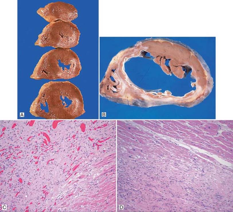
Fig 2 Myocardial infarction. Heart slices demonstrate large extent of an old, scarred infarct along the left ventricular free wall (A). Note the prominent left ventricular hypertrophy. Single cross-section of the ventricles shows healed myocardial infarct involving the interventricular septum and anterior and posterior left ventricular walls (B). Microscopically, a healing infarct of about 2 weeks of age will show some fibrosis and granulation tissue (C).Infarcts more than 2 months old will show dense fibrosis adjacent to normal-appearing myocardium (D).

Fig 3 Myocardial infarction. Papillary rupture, a complication of acute MI. Note the tissue surrounding the rupture is soft and darkened (A). Left ventricular aneurysm, another complication, is seen in B.