
Heart transplantation is performed in both pediatric and adult populations
Indications
• Systolic heart failure (left ventricular ejection fraction, <35%) due to ischemic cardiomyopathy, dilated cardiomyopathy, and hypertensive and valvular diseases
• Ischemic heart disease with intractable angina not amenable to other therapies
• Intractable arrhythmias not amenable to an implantable cardioverter and defibrillator
• Hypertrophic cardiomyopathy
• Congenital heart diseases in which fixed pulmonary hypertension is not a complication
Exclusions
• Amyloidosis
• HIV infection
• Malignancy, other than basal cell carcinoma
Examination
• Weigh the heart
• Examine and describe epicardial surfaces
• All chambers should be stuffed with formalin-soaked gauze and fixed in formalin overnight
• Examine coronary arteries in situ by cross-sectioning at every 0.3- to 0.5-cm intervals
• For ischemic heart disease: serially section transversely (bread loaf) 1 cm thick beginning at the apex of the heart and extending to the level of chordae tendinae of the mitral valve
• For nonischemic cardiomyopathy (dilated or hypertrophic): bisect longitudinally from apex to base, bivalving both ventricles and bisecting tricuspid and mitral valves (apical four-chamber cut)
• For congenital heart disease: cut heart along lines of blood flow (inflow–outflow)
• Paraffin sections:
• Cross-sections of each major coronary artery
• Three from left ventricle, apex, mid, and base
• Two from interventricular septum
• Two from right ventricle
• Sample any valvular or myocardial lesion
Outcomes
• About 90% survival rate at 1 year
• About 80% survival rate at 5 years
• About 70% survival rate at 10 years
• Survival is better in infants who receive a transplant in the first year and survive the early posttransplant period (i.e., after the first year of life)
Complications
• Surgical: rarely seen now with increased experience and better techniques; include early graft failure, bleeding, wound infection, stenosis of vascular anastomosis
• Rejection
• Antibody-mediated
• Acute cellular
• Chronic rejection (accelerated graft vasculopathy)
• Infection: typically the grafted heart is not involved; bacterial and opportunistic infections occur in lungs and gastrointestinal tract
• Posttransplant lymphoproliferative disease: incidence has decreased to less than 3%
• Perforation during endomyocardial biopsy: histologically, a fragment or fragments of epicardium consisting of adipose tissue and mesothelial (calretinin-positive) cells can be seen
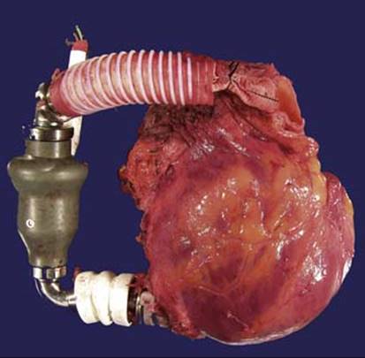
Fig 1 Heart transplantation. Sometimes a heart is received with hardware such as left ventricular assist device that needs to be taken off before gross examination.
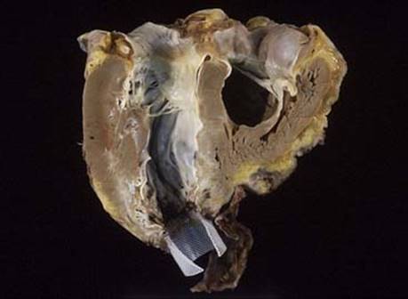
Fig 2 Heart transplantation. Hypertrophic cardiomyopathy with a four-chamber cut and part of left ventricular assist device still attached.
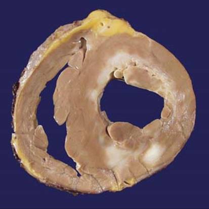
Fig 3 Heart transplantation. Ischemic heart disease: bread loaf cut of this heart shows focal areas of scarring.
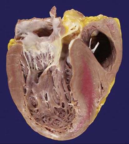
Fig 4 Heart transplantation. Dilated cardiomyopathy (four-chamber cut); as often seen in transplantation, portions of atria are missing in the explanted heart. Also note poor fixation of the interventricular septum.
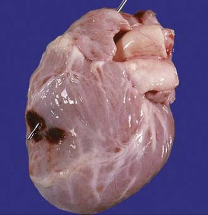
Fig 5 Heart transplantation. An unusual complication: endomyocardial biopsy perforation that led to cardiac tamponade and this child’s death.
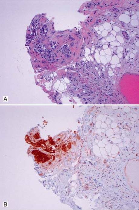
Fig 6 Heart transplantation. This endomyocardial biopsy contains a fragment of epicardial fat lined by mesothelial cells (A), which are confirmed by positive calretinin stain (B).