
Definition
• An accelerated form of coronary disease with uniform intimal fibrosis involving both epicardial and intramural coronary arteries and veins
Clinical features
Epidemiology
• Incidence of CAV increases with time in both pediatric and adult patients, being less in the former
• In pediatric patients the incidence is 2.5% at 1 year, 11% at 5 years, and 16.7% at 10 years after transplantation
• In adults, the incidence is 8% at 1 year, 30% at 5 years, and approximately 50% at 10 years after transplantation
Presentation
• May be seen as ventricular arrhythmias, congestive heart failure, or sudden death
Prognosis and treatment
• Augmented immunosuppression
• Coronary artery bypass graft
• Percutaneous coronary intervention
• The only definitive treatment is retransplantation, but results are poor
Pathology
Histology
• The whole length of the coronary vessels is usually affected
• Involvement of large epicardial vessels shows concentric intimal proliferation composed of smooth muscle cells and less differentiated spindled cells (myofibroblasts or “myointimal” cells)
• Lesions in the small intramyocardial branches are characterized by diffuse concentric narrowing with luminal stenosis due to intimal fibrosis
Immunopathology/special stains
• Not contributory
Main differential diagnosis
• Preexisting coronary atherosclerosis: typically eccentric plaques with cholesterol clefts, foamy macrophages, and calcifications
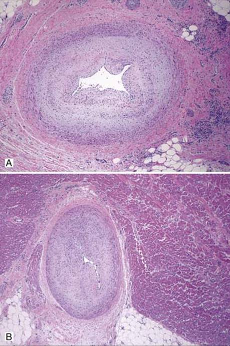
Fig 1 Chronic rejection of the heart. These low-power views show epicardial arteries with concentric intimal fibrosis with near-complete luminal obliteration (A and B).
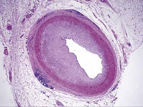
Fig 2 Chronic rejection of the heart. Intimal fibrosis of coronary artery may be eccentric, as seen here. Also note the presence of chronic inflammation in the adventitia, which is often seen in CAV.
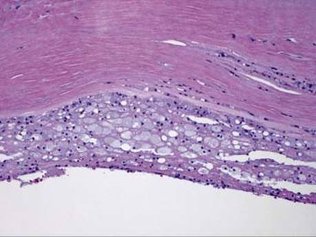
Fig 3 Chronic rejection of the heart. This example of CAV has foamy macrophages and intimal fibrosis, which raises the differential diagnosis of preexisting atherosclerosis in the transplanted heart.
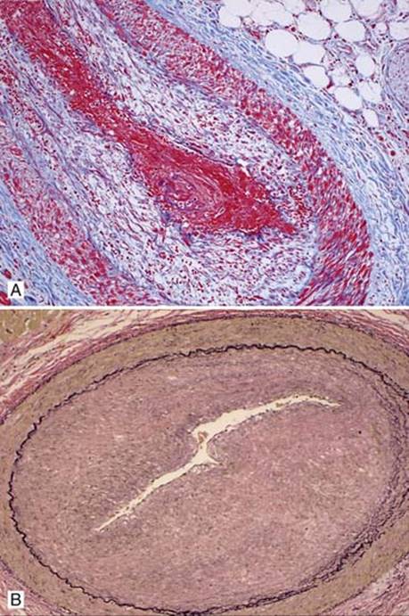
Fig 4 Chronic rejection of the heart. Trichrome (A) and elastic (B) stains highlight intimal fibrosis. Note minimal elastic layer disruption that is characteristic in CAV.
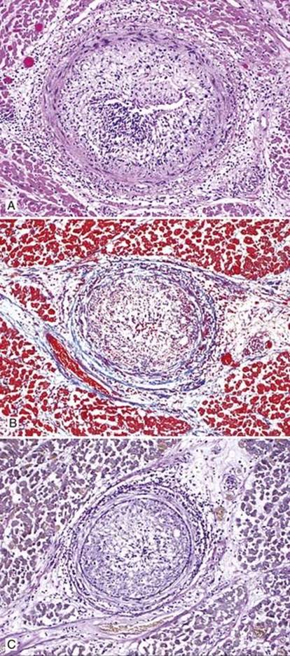
Fig 5 Chronic rejection of the heart. Intramyocardial coronary artery with marked intimal fibrosis and significant inflammatory infiltrates (mostly lymphocytes), H&E (A), trichrome (B), and EVG (C)stains.