
Definition
• Acute cellular rejection occurs from days to years after transplantation and is characterized by infiltrates of activated lymphocytes, macrophages, and occasional eosinophils
Clinical features
Epidemiology
• Can occur at any time from days to months or years after transplantation
• At least 80% of patients experience at least one episode of rejection
Presentation
• Usually asymptomatic because patients are given a diagnosis at time of biopsy
• Only approximately 5% of patients exhibit severe hemodynamic compromise
Prognosis and treatment
• Treatment varies and may include oral and intravenous corticosteroids, antithymocyte globulin, and/or murine monoclonal antibody OKT-3
• Most patients recover from acute rejection
• Repeated episodes of acute rejection increase the risk of accelerated graft vasculopathy
Pathology
Histology
• Endomyocardial biopsies are graded using the International Society of Lung and Heart Transplantation nomenclature published in 1990 and revised in 2005. A comparison of the two grading systems is provided in Web Table 5on the Expert Consult website
• Focal mild rejection (G1A/G1R):
• Focal perivascular interstitial infiltrate consisting of activated lymphocytes
• Infiltrates usually seen in one level or become smaller on subsequent levels
• Diffuse mild rejection (G1B/G1R):
• Few activated lymphocytes between myocytes with no eosinophils or muscle damage
• Focal moderate rejection (G2/G1R):
• Can be single infiltrates often containing eosinophils and macrophages and few plasma cells with focal myocyte damage
• Multifocal moderate rejection (G3A/G2R):
• Two or more foci of infiltrate with associated myocyte damage
• Severe rejection (G3B and G4/G3R):
• Rarely seen with current regimens and is manifested by diffuse aggressive infiltrates (lymphocytes, eosinophils, and neutrophils) with myocyte necrosis and damage
Immunopathology/special stains
• To exclude a Quilty lesion, an immunostain for CD21 can be helpful. CD21 stain is positive in the center of large Quilty lesions and is negative in rejection
Main Differential Diagnoses
• Infection: mixed inflammatory infiltrates
• Quilty effect: is an endocardial infiltrate that is not associated with acute cellular rejection and often infiltrates into the underlying myocardium
• Site of previous biopsy:
• Often seen in the first months after transplantation
• Caused by the fact that biopsy catheter tracks to same region
• Organizing fibrin and granulation tissue
• Reactive endothelial cells and macrophages
• Rare inflammatory cells present
• Later stages demonstrate fibrosis

Fig 1 Acute cellular rejection of the heart. This low-power view show an essentially normal endomyocardial biopsy with no cellular infiltrates (grade G0).
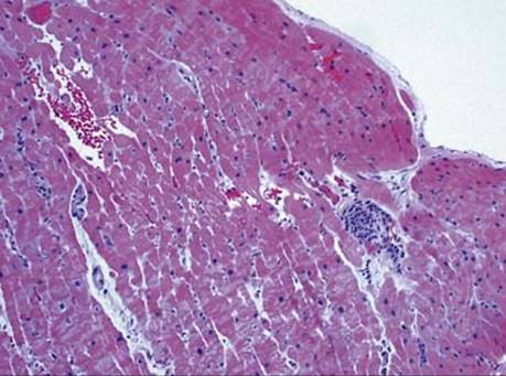
Fig 2 Acute cellular rejection of the heart. This low-power view show a small focus of perivascular interstitial infiltrate consisting of activated lymphocytes (grade G1A/1R).
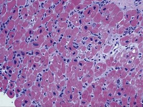
Fig 3 Acute cellular rejection of the heart. This high-power view shows an interstitial infiltrate consisting of few activated lymphocytes between myocytes with no eosinophils or muscle damage (grade G1B/1R).

Fig 4 Acute cellular rejection of the heart. This medium-power view shows an area of lymphocytic infiltrate with associated muscle damage (grade G2/1R).

Fig 5 Acute cellular rejection of the heart. There are extensive interstitial lymphoid infiltrates (grade 3B/3R) with associated muscle damage: low (A) and high (B) powers.

Fig 6 Acute cellular rejection of the heart. Site of previous biopsy with organizing fibrin and few inflammatory cells.
WEB TABLE 5 Comparison of 1990 and 2005 Heart Rejection Grading Formulations
|
DESCRIPTION OF REJECTION |
1990 GRADE |
2005 GRADE |
|
Focal mild, mild and focal moderate |
1A, 1B, 2 |
1R |
|
Multifocal moderate |
3A |
2R |
|
Diffuse moderate, severe |
3B, 4 |
3R |