
Definition
• Benign tumor(s) of cardiomyocytes that have abundant vacuolated cytoplasm; now considered to be hamartoma
Clinical features
Epidemiology
• The most common cardiac tumor in infants
• Usually diagnosed in children less than 1 year of age
• Half of the patients have tuberous sclerosis (TS) with mutations in TSC1 and TSC2 genes
• Sporadic cases may be associated with congenital heart disease
• No known sex predilection
Presentation
• May be asymptomatic
• Can be found during prenatal period by routine ultrasound examination detecting a mass or associated nonimmune hydrops or fetal cardiac arrhythmias
• Rhabdomyomas are seen early in life with obstruction of the inflow/outflow pathways (cardiac valve or chamber) or arrhythmias due to disruption of the nodal or septal conduction system by the tumor or sudden death
• Rhabdomyomatosis: numerous miliary nodules less than 1 mm
Prognosis and treatment
• High rate of spontaneous regression in young children, especially when associated with TS; less likely to regress in older children
• Complete surgical resection is curative
• Despite complete resection, risk of sudden death remains due to damage to the conduction system
Pathology
Gross
• Small intramural tumors; commonly multiple; most often involving the left ventricle followed by the right
• Usually small, firm, gray-white, well-circumscribed nodules protruding into the ventricles
• Approximately 3 cm to 4 cm in size; however, can grow to up to 10 cm in sporadic cases
Histology
• Composed of clear cells and large, round, polygonal cells termed spider cells with glycogen vacuoles separated by strands of cytoplasm extending between the cell membrane and nucleus
• Cross-striations are identifiable in the cytoplasm
• No mitotic activity
• Adult rhabdomyomas are more cellular and have smaller cells and few spider cells
Immunopathology/special stains
• PAS positive and diastase sensitive (glycogen)
• Positive for myoglobin, actin, desmin, vimentin; variable HMB45
• Negative for S100
Main differential diagnoses
• Lipoma: epicardial; no spider cells, glycogen or myofibers
• Granular cell tumor: epicardial; no vacuoles or myofibers; S100 positive; desmin and myoglobin negative
• Glycogen storage disorder
• Histiocytoid cardiomyopathy: finely granular cells; no large vacuoles; no spider cells
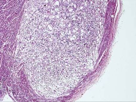
Fig 1 Cardiac rhabdomyoma. The tumor is seen protruding into the ventricular cavity, covered by a thin rim of compressed myocytes and endocardium.
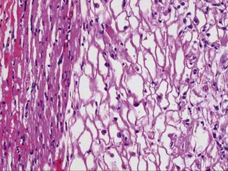
Fig 2 Cardiac rhabdomyoma. High-power image of rhabdomyoma shows that it is composed of clear cells that stand in contrast to normal cardiomyocytes on the left.
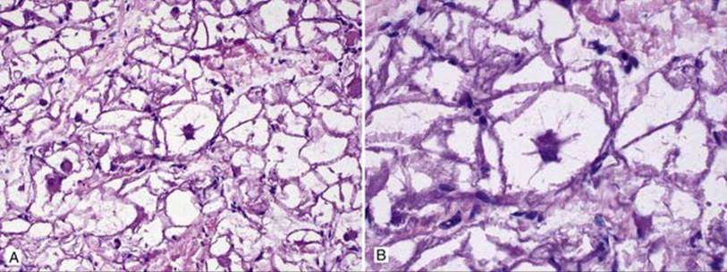
Fig 3 Cardiac rhabdomyoma. Among the clear cells are few large “spider cells” with glycogen vacuoles that are separated by strands of cytoplasm seen at low (A) and high (B) power.
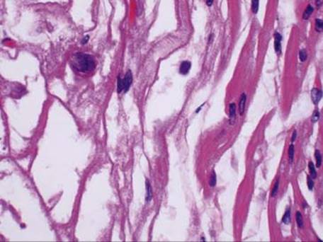
Fig 4 Cardiac rhabdomyoma. Cross-striations are seen within the tumor cells on highest power.
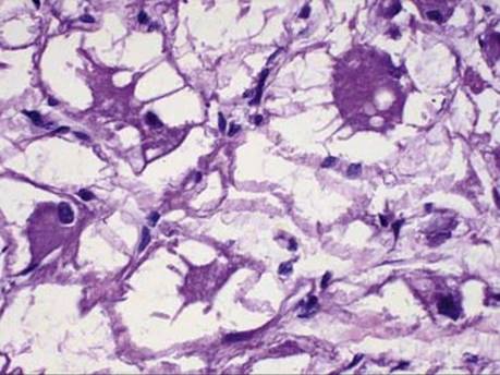
Fig 5 Cardiac rhabdomyoma. High-power image showing that the intracellular glycogen is pushing the cytoplasm but not the nucleus.