
Definition
• A benign, papillary lesion of endocardial origin lined by endothelial cells and a core composed of fibrous tissue and elastic stroma
Pathogenesis
• Etiology largely unknown, and few pathogenic mechanisms have been proposed
• Some theories include a neoplastic proliferation or a hamartoma
• Others have described the lesion as a response to trauma, cardiac injury, or focus of disease
• Evidence suggests that a subset of lesions arise in areas of diseased valves (e.g., mitral valve prolapse, rheumatic heart disease, and atrial septal defect); hemodynamic changes in areas of disease may lead to exuberant response
• Another study described incidence of lesion in areas of previous iatrogenic interventions (e.g., cardiac surgery or chest irradiation)
• Others have found cytomegalic changes in some cases, leading to the belief that it may represent a chronic viral endocarditis
Clinical features
Epidemiology
• It is the second most common primary tumor of the heart after myxoma, but comprises only 10% of all primary cardiac neoplasms
• About 90% of cardiac fibroelastomas occur on the valves (aortic>mitral>>tricuspid and pulmonic); may also occur on papillary muscles and chordae
• It is the most common primary tumor of cardiac valves and can occur anywhere on the valve (as opposed to Lambl excrescences—see further on)
• Patients are often middle-aged to elderly adults, but it can affect all ages
Presentation
• Patients may present with varying symptoms depending on the location of the lesion; some may be asymptomatic
• Lesions near the ostia of vessels may manifest with angina, myocardial infarction, or even sudden death
• Papillary fronds of the tumor may slough off, leading to distal emboli, most often in cerebral vessels that manifest as strokes
• Damage to endothelial covering leads to thrombi formation, which can also embolize
• Clinical evaluation includes transthoracic or transesophageal echocardiograms; chest CT may also be performed
Prognosis and treatment
• Most cases are treated with surgical excision and have a good overall prognosis
Pathology
Gross
• The lesion is a small (usually <1 cm) papillary, somewhat gelatinous, white-tan, pedunculated, often mobile mass arising most often from a valve or the endocardium
• They most often occur singly, but in cases arising from iatrogenic effects, they can be multiple
• The multiple fronds, when the tumor is placed in water, are likened to a sea anemone
Histology
• The papillary appearance of the lesion is readily appreciable; the branching fronds are lined by endothelial cells overlying a proteoglycan-rich layer of tissue (intermediate layer)
• The central core of the fronds is avascular and contains stroma rich in elastic fibers
• Inflammatory cells may be present within the core, including dendritic cells
• Complicated lesions may demonstrate thrombi formation
Immunopathology/special stains
• The surface lining cells immunostain for endothelial cell markers, including CD31, CD34, and factor VIII
• The intermediate layer stains positive for vimentin and focally for S100 protein (probably dendritic cells)
• The central layer has positivity for elastin
Main differential diagnoses
• Lambl excrescences: often smaller and occur only on the midline of the sites of valve closure; not as gelatinous; no elastic tissue in core
• Myxoma: no elastic tissue in core; cells are polygonal and have larger nuclei and more abundant cytoplasm
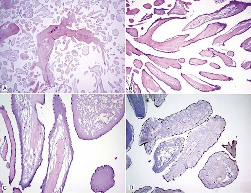
Fig 1 Cardiac fibroelastoma. Low- (A), medium- (B), and high- (C) power views show a papillary lesion with numerous, branching fronds, some of which have acellular pink material in their cores. D,CD31 immunostain highlights endothelial cells covering the papillae.

Fig 2 Cardiac fibroelastoma. Elastic stain highlights elastic fibers in some of the cores, low (A) and high (B) powers.