
Definition
• Benign proliferation of blood vessels
Clinical features
Epidemiology
• Rare in the heart
Presentation
• Often incidental finding
• May cause arrhythmias, syncope, or sudden death
Prognosis and treatment
• Excision is curative
Pathology
Gross
• May be well-circumscribed or infiltrating
• Intramyocardial or subendocardial mass protruding into the atrium or ventricle
Histology
• Capillary, cavernous, or arteriovenous patterns can be seen
• May have myxoid background
• Intramural (intramyocardial) tumors frequently have an arteriovenous pattern and infiltrative borders and are associated with fibrous tissue and fat
• Papillary endothelial hyperplasia can occur
Immunopathology/special stains
• Vascular markers are positive but not usually needed for diagnosis
Main differential diagnoses
• Angiosarcoma: foci of necrosis, nuclear atypia, and high mitotic activity
• Myxoma: stellate (myxoma) cells, intermixed inflammatory cells, and hemosiderin-laden macrophages
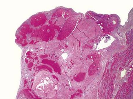
Fig 1 Cardiac hemangioma. Right atrial hemangioma with cavernous morphological features. Some of the vascular spaces contain organizing thrombi.
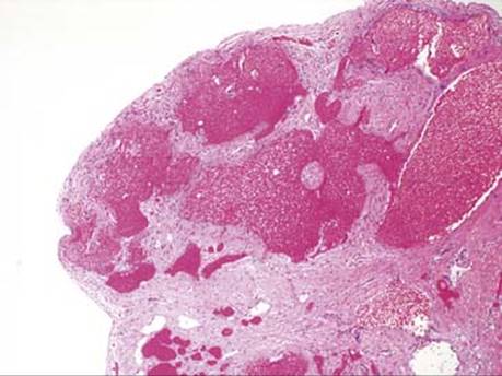
Fig 2 Cardiac hemangioma. Higher power of lesion seen in Fig 1.

Fig 3 Cardiac hemangioma. Left ventricular papillary muscle hemangioma, polypoid, with primarily capillary morphological features.

Fig 4 Cardiac hemangioma. Low-power view of same case as in Fig 3 shows thin-walled vessels that focally invade the underlying myocardium.
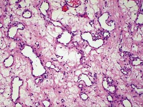
Fig 5 Cardiac hemangioma. Myxoid degeneration can occur (high-power view of Fig 3).
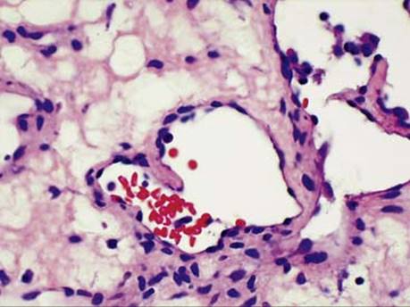
Fig 6 Cardiac hemangioma. The nuclei are bland, and mitoses are not apparent (high-power view of Fig 3).

Fig 7 Cardiac hemangioma. Compare to low-power view of organizing thrombus attached to endocardium. Myocardium is on the left.

Fig 8 Cardiac hemangioma. Medium power of Fig 7 showing small blood vessels growing into the thrombus.

Fig 9 Cardiac hemangioma. High power of Fig 7 showing proliferating endothelium with poorly slit-like vascular spaces.