
Definition
• Rare benign neoplasm of cardiac muscle, seen as a single mass commonly involving the interventricular septum or left ventricular free wall
Clinical features
Epidemiology
• Second most common cardiac tumor most frequently seen in children younger than 1 year of age
• Sudden death in one third of the cases is caused by arrhythmias or outflow obstruction
• No known sex predilection
• Unknown etiology but with causal association with Gorlin syndrome (syndrome of multiple nevoid basal cell carcinomas, cysts and fibrosarcomas of the jaws, and skeletal abnormalities; multiple skin abnormalities, including milia, epidermoid cysts, chalazia, and comedones; associated with medulloblastoma, meningioma, ovarian fibroma/fibrosarcoma, cardiac fibroma, fetal rhabdomyoma, and mesenteric lymphatic or chylous cysts)
Presentation
• Presents with heart failure, cyanosis, syncope, or arrhythmias
• Symptoms are related to impairment of left ventricular wall function with reduction in left ventricular filling capacity and ejection fraction
• Arrhythmias leading to sudden death and abnormalities of atrioventricular conduction are due to disruption of the nodal or septal conduction system by the tumor
• Small cardiac fibromas can be asymptomatic and found incidentally on cardiac studies while other cardiac disease is being investigated
• Tumor embolization is uncommon
Prognosis and treatment
• Fibromas do not regress; they gradually grow larger
• Complete surgical resection is curative, when possible
• Despite complete resection, risk of sudden death remains due to damage to the conduction system
• If the tumor is too large to resect (can occasionally exceed 10 cm), cardiac transplantation might be considered
Pathology
Gross
• Discrete solitary bulging mass with calcifications commonly involving the interventricular septum or left ventricular free wall
• Minority of reported cases with atrial or great vessel involvement
• Firm, trabeculated cut surface resembling leiomyomas
Histology
• Bland fibroblasts and collagen fibers with infiltrative margins and calcification
• Perivascular lymphocytes, histiocytes, elastic fibers, and myxoid stroma are occasionally seen
• More cellular with mitotic figures in infants
• Calcification and cystic degeneration in older children
Immunopathology/special stains
• Both smooth muscle actin (SMA) and desmin stain compressed cells between the thick collagen bundles
Main differential diagnoses
• Fibrosarcoma: rare in young children; mitotically active
• Inflammatory pseudotumor/myofibroblastic tumor: endocardial lesions covered by fibrin with stellate tumor cells with prominent nucleoli, more inflammation, occasional mitosis, and necrosis
• Scars
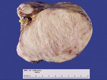
Fig 1 Cardiac fibroma. This large well-circumscribed tumor was removed from a 64-year-old woman. It measured 12.7 cm in largest dimension. The cut surface was firm and fibrous-appearing.

Fig 2 Cardiac fibroma. Low-power view shows the well-circumscribed tumor consisting of dense fibrous tissue adjacent to cardiac muscle.
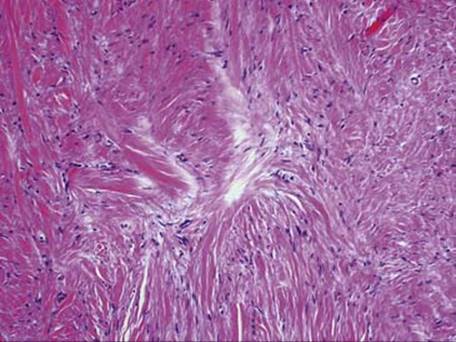
Fig 3 Cardiac fibroma. High power shows dense fibrous tissue with bland fibroblasts and thick collagen fibers in the tumor.

Fig 4 Cardiac fibroma. Thin-walled vessels are present within the dense fibrous tissue of the tumor.

Fig 5 Cardiac fibroma. SMA stains compressed cells between the dense collagen fibers.