
Definition
• Deposition of fat in the atrial septum at the level of the fossa ovalis
• Approximately 2 cm or greater in width
Clinical features
Epidemiology
• Usually discovered in older, obese patients
• Rarely associated with metabolic syndromes
• Incidental finding in 1% to 8% of the population
Presentation
• Most commonly an incidental finding
• Rarely associated with arrhythmias or superior vena cava (SVC) obstruction
Prognosis and treatment
• Surgical excision only for symptomatic SVC obstruction or intractable arrhythmias
• Rare cause of sudden death due to arrhythmias
Pathology
Gross
• Unencapsulated fatty mass often extending into the right atrium
• Thickness superior to the fossa ovalis is greater than the thickness inferior to the fossa ovalis
• Fossa ovalis is usually spared, resulting in a “dumbbell” appearance
• Often continuous with subepicardial adipose tissue
Histology
• Infiltrating adipocytes between enlarged, often bizarre cardiac myocytes
• At least some adipocytes are multivacuolated, similar to brown fat
• No lipoblasts or mitotic figures are present
• Bands of collagen or inflammatory cells may be present
Immunopathology/special stains
• Not contributory
Main differential diagnoses
• Liposarcoma: contains lipoblasts with enlarged, indented nuclei and mitotic figures
• Lipoma: encapsulated with no vacuolated adipocytes
• Myxoma: myxoid stroma; no hypertrophic myocytes or adipocytes

Fig 1 Lipomatous hypertrophy of the interatrial septum. Thickening of the interatrial septum by nonencapsulated fatty tissue (arrow).

Fig 2 Lipomatous hypertrophy of the interatrial septum. Adipocytes infiltrating between cardiac myocytes with enlarged, hyperchromatic nuclei and associated fibrosis and inflammation.
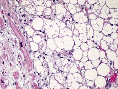
Fig 3 Lipomatous hypertrophy of the interatrial septum. Adipocytes with multiple vacuoles similar to brown fat adjacent to hypertrophic cardiac myocytes.