
Definition
• Malignant proliferation of endothelial cells
Clinical features
Epidemiology
• Most common primary cardiac sarcoma
• More frequent in males
• Average age, 40 years
Presentation
• Chest pain, hemopericardium, tamponade, or outflow obstruction
• Frequent metastases at time of presentation (most commonly to the lung)
Prognosis and treatment
• Standard treatment involves resection, radiation therapy, and chemotherapy; complete resection is rarely possible
• Average survival is less than 1 year
Pathology
Gross
• Most commonly occurs in the right atrium and may extend into veins, through tricuspid valve and through pericardium
• Multilobular tumor
Histology
• Anastomosing vascular channels lined by atypical cells, often with papillary tufts
• May have cytoplasmic vacuoles containing red blood cells, which is a helpful feature for diagnosis
• Large areas of undifferentiated spindled cells are frequently present
• Areas of benign-appearing vascular channels are common
• Frequent mitotic figures and necrosis
Immunopathology/special stains
• CD31 has the highest sensitivity of vascular markers but may be weak, focal, or negative
• CD34 and factor VIII–related antigen may also be positive but are less sensitive
Main differential diagnoses
• Undifferentiated spindle cell sarcoma: no foci of vascular channels
• Kaposi sarcoma: most common in immunosuppressed patients; frequently seen as multiple small nodules and positive for human herpesvirus 8
• Hemangiomas: can have papillary tufts of endothelial cells but no necrosis or solid foci of spindle cells
• Metastases: clinical history
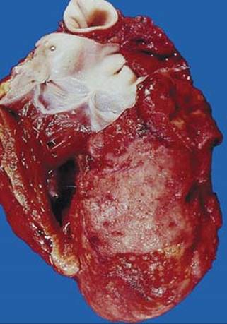
Fig 1 Angiosarcoma of the heart. Angiosarcoma at autopsy extended to right ventricle.

Fig 2 Angiosarcoma of the heart. This angiosarcoma has a solid spindle-cell component, but typical anastomosing vascular channels are present in the lower left.

Fig 3 Angiosarcoma of the heart. Large, atypical cells project into the vascular space.

Fig 4 Angiosarcoma of the heart. In an undifferentiated area of this angiosarcoma, red blood cells are seen within intracytoplasmic lumen.

Fig 5 Angiosarcoma of the heart. CD31 is strongly positive in this angiosarcoma.

Fig 6 Angiosarcoma of the heart. In this biopsy of apparently undifferentiated spindled cells, immunohistochemical tests for vascular markers would be required for diagnosis.