
Definition
• Secondary malignant tumor of the heart, arising from lymphatic or hematogenous spreading of a primary neoplasm
Pathogenesis
• Esophageal, lung, and breast carcinomas metastasize to the mediastinal lymph nodes and invade the lymphatics of the epicardium and pericardium. May be superficial myocardial involvement
• Melanomas, lymphomas/leukemias, sarcomas, renal cell carcinomas, and choriocarcinomas have a marked tendency to spread hematogenously and involve the myocardium
• Malignancies in adjacent organs, such as lung or esophageal carcinomas, may reach the heart by direct extension
• May be extension of tumor through intraluminal growth in the draining veins to the vena cava and right atrium (e.g., hepatocellular carcinoma, renal cell carcinoma, and Wilms tumor). Lung carcinomas may reach the heart via extension in the pulmonary vein
Clinical features
Epidemiology
• Metastatic lesions in the heart are more common than primary tumors, by a factor of about 30 to 1
• Virtually any tumor can metastasize, but the most common sources include lung, breast, kidney, and thyroid carcinomas and melanomas. Lymphomas and leukemias may also involve the heart
• Rate of cardiac metastasis in a number of autopsy series is about 12% to 25% in patients with widespread malignancy
• Generally, the right side of the heart is involved in one third of cases, the left side is involved in another one third, and bilateral disease is present in the remaining one third
• Usually there are multiple metastatic tumor deposits; single nodules are rare but these may be surgically resectable
Presentation
• Presentation is variable depending on location of tumor deposit(s)
• Involvement of pericardium may lead to pericardial effusions
• Myocardial involvement may lead to infarction (creating a diagnostic dilemma), conduction defects and arrhythmias, and congestive heart failure (with dyspnea, hypoxia, and hypotension)
• Disease in valves may lead to obstruction with syncope
Prognosis and treatment
• Cardiac metastases are often missed during work-up of malignancies, may not be detected until years after the primary diagnosis, and are often not detected at all until autopsy
• Echocardiography is the method of choice for detecting cardiac involvement
• Pericardiocentesis may be performed if effusion is present
• In most cases, the heart is not exclusively involved; metastatic disease is widespread when cardiac involvement is detected
• Treatment may involve pericardiocentesis to relieve the effusion. Surgical resection of single nodules may be performed if indicated
Pathology
Gross
• Pericardial metastasis may result in a desmoplastic reaction, leading to constrictive pericarditis, usually due to extension of a pulmonary malignancy. Diffuse studding and thickening of the pericardium may also occur
• Myocardial metastases on cut section demonstrate nodular, white-tan infiltrates throughout
• Less common is a single, dominant mass or an intracavity lesion
Histology
• Microscopic sections will demonstrate morphological features of the primary malignancy
Immunopathology/special stains
• Immunohistochemical analysis may be helpful if the primary tumor is unknown or if it was diagnosed many years ago
• Morphological features will, in most cases, be similar to the primary malignancy
• In cases of high-grade, undifferentiated tumor, a preliminary panel should include keratins and melanocytic and lymphoid markers
Main differential diagnoses
• Malignant mesothelioma: calretinin, WT-1, D2-40 and CK5/6 positive; MOC31 and BG8 negative
• Primary cardiac tumor: usually single lesion with characteristic histological features
• Myocardial infarction: may be a consideration at autopsy; histological analysis will be diagnostic
• Valvular disease: metastasis on valves may mimic vegetations; histological analysis will be diagnostic

Fig 1 Cardiac metastasis. Metastatic breast carcinoma extending into myocardium.
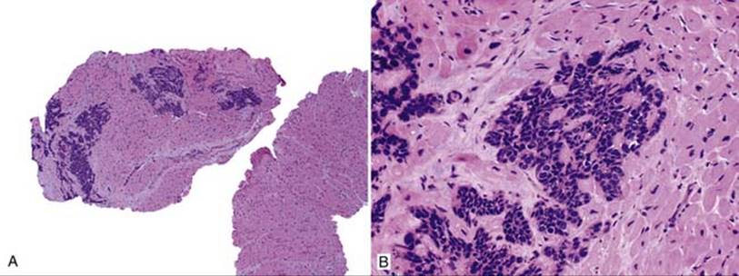
Fig 2 Cardiac metastasis. Endomyocardial biopsy with infiltration of cardiac myocytes by metastatic squamous cell carcinoma: low (A) and high (B) powers.
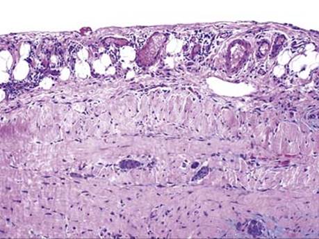
Fig 3 Cardiac metastasis. Pericardial biopsy with metastatic lung adenocarcinoma.
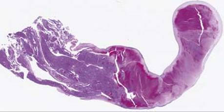
Fig 4 Cardiac metastasis. This left atrial tumor with attached thrombus was pulled out from a 49-year-old with metastatic malignant melanoma.
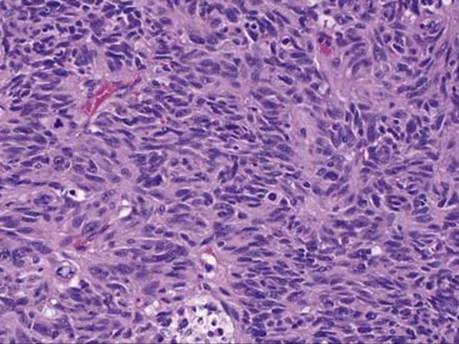
Fig 5 Cardiac metastasis. High-power view of the tumor in Fig 4 shows spindle cells with mitoses.
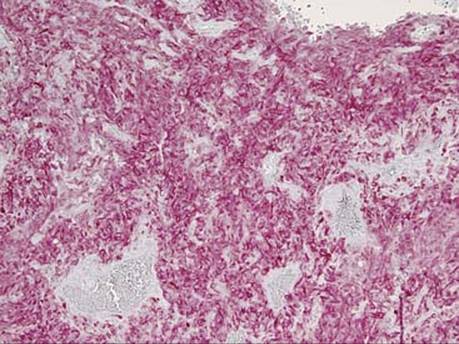
Fig 6 Cardiac metastasis. The tumor shown here is positive for Melan-A (it was also positive for S100 and HMB45), confirming melanocytic origin.