
Definition
• Vascular disease characterized by formation of an atheroma in various arteries due to endothelial injury and healing, leading to plaque formation
Pathogenesis
• Lesions begin as fatty streaks in childhood with lipid-laden macrophages under endothelium
• Initial injury thought to be due to a variety of factors, including hypertension, hypercholesterolemia/hyperlipidemia, smoking, elevated homocysteine levels, various toxins, and/or certain viruses
• Hemodynamic factors and hypercholesterolemia are the two most important causes of endothelial injury. Areas of vessel wall without turbulent flow have endothelial protein expression of factors that protect against oxidant damage and atheroma formation. Also adhesion molecules and inflammatory proteins are upregulated in areas with turbulent flow (such as ostia of branch vessels)
• Hypercholesterolemia causes infiltration of low-density lipoprotein (LDL) into the intima, leading to production of oxygen free radicals and oxidized LDL, which is not only cytotoxic but also allows production of cytokines and growth factors, initiating an inflammatory cascade
• Monocytes and T cells migrate into the intima; macrophages engulf oxidized LDL (becoming foam cells) and are activated, releasing more cytokines and growth factors that results in smooth muscle cell migration from the vessel wall media. Platelets also infiltrate and are activated
• Smooth muscle migration and proliferation occurs; these cells produce a collagen-rich matrix, which forms a fibrous cap over the underlying atheroma
• Erosion or ulceration exposes a highly thrombogenic surface that can result in acute thrombosis of the vessel
• Rupture of the plaque may also lead to distal microemboli
Clinical features
Epidemiology
• Atherosclerotic disease is endemic in the western world, where it is the leading cause of death; it is also common in Southeast Asia; prevalence rates are lower in Latin America, Africa, and the Far East
• Most commonly involves the lower abdominal aorta, coronary arteries, popliteal arteries, and the circle of Willis
• It is responsible for about half of all deaths in the United States from associated conditions such as ischemic heart disease and cerebrovascular disease; 90% of deaths due to heart disease are attributed to atherosclerosis
• Major risk factors include age (uncommon before fourth decade), gender (hormonal protection in premenopausal women), family history, hyperlipidemia (especially high cholesterol), hypertension, diabetes, and smoking
• Other risk factors include obesity, hyperhomocysteinemia, coagulation disorders, sedentary lifestyle, and a high-stress environment
Presentation
• Atherosclerotic disease is one of the “silent killers” whose progression often goes unnoticed until late in the disease course
• Stable angina can result from fixed stenosis of coronary vessels (leading to episodic ischemia of myocardium) and is manifested by periods of chest pain or discomfort during exercise or emotional excitement; unstable angina results from acute disruption of the atheroma and is manifested by severe, progressively worsening chest pain that occurs even at rest
• Acute changes in the plaque can lead to myocardial infarction; these acute changes may arise in a mild to moderately stenotic vessel, not necessarily in a severely occluded lesion
• Involvement of peripheral vessels results in claudication in lower extremities
• Progressive atherosclerotic disease in cerebral vessels can lead to transient ischemic attacks and later ischemic strokes
Prognosis and treatment
• With presence of risk factors, untreated fatty streaks can progress to fibrofatty plaques; with continued inflammation, thrombi formation and calcification occur; these become advanced lesions
• Advanced lesions are vulnerable to complications: vessel occlusion (due to plaque growth), thrombosis (due to plaque rupture and exposure of underlying thrombogenic surface), hemorrhage (due to rupture of thin-walled vessels of neovasculature), and aneurysms/dissections or vessel rupture (due to wall weakening secondary to ischemic atrophy of media and loss of elastic fibers)
• Treatment involves control of modifiable risk factors, including lowering cholesterol (e.g., with statins), controlling hypertension and blood sugar levels, losing weight, and stopping smoking; aspirin is often prescribed to prevent platelet activation at the site of a potentially thrombogenic lesion
• Discovery that disease has an inflammatory nature has led to progress in developing vaccines and antiinflammatory drugs
Pathology
Gross
• Fatty streaks represent the coalescence of small, yellow flat spots near the ostia of branch vessels
• Atheromatous plaques appear as thickened, hardened 0.5-cm to 1.5-cm foci of the vessel wall; ulceration is often seen. Lesions may be at various stages of development within the same vessel
• Complicated lesions may show rupture of plaque or areas with thrombosis and hemorrhage into the plaque
• Atherosclerotic aneurysm (see next chapter)
Histology
• Typically, partial or complete occlusion of the vessel lumen occurs with an eccentric lesion that is “capped” at one end by a fibrous band composed of smooth muscle cells and collagen. Beneath this is a necrotic core containing foam cells, cholesterol crystals, and debris from dead cells. At the periphery is a collection of inflammatory cells (macrophages and T cells) along with thin-walled vessels formed by neovascularization
• Calcification and neovascularization are common
• Some lesions show different proportions of smooth muscle cells, extracellular matrix, and lipid
• Histological sections of acute plaque change show disruption of the fibrous cap with or without subsequent thrombus formation over it; this may completely occlude the vessel
Immunopathology/special stains
• Trichrome stain highlights fibrous cap
• Elastic stain shows loss of fibers in the media
Main differential diagnosis
• None
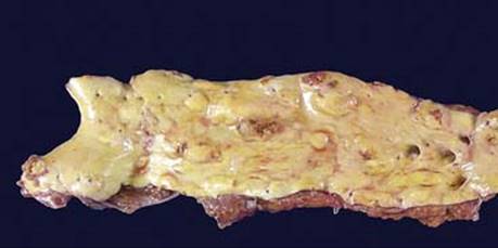
Fig 1 Atherosclerosis. Gross photograph of section from abdominal aorta showing numerous, slightly elevated, yellow-tan plaques distributed throughout the vessel wall.
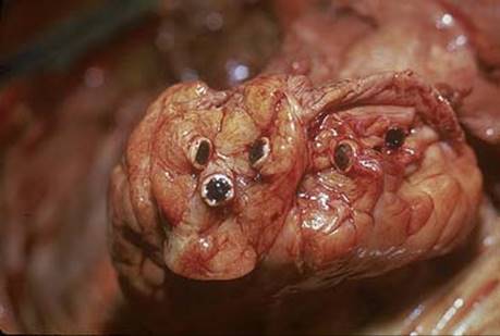
Fig 2 Atherosclerosis. Coronary vessels demonstrating a feared complication of atherosclerosis: acute plaque change with hemorrhage, leading to acute vessel thrombosis and distal myocardial infarction.
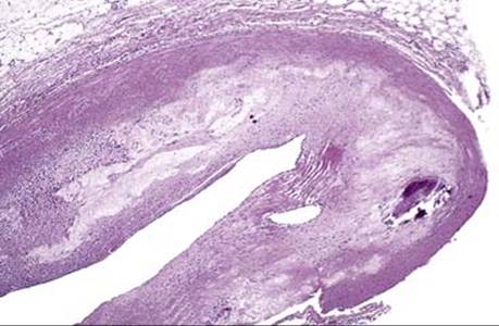
Fig 3 Atherosclerosis. Low-power view of an atherosclerotic lesion, demonstrating a fibrous cap and a necrotic core that compromises the vessel lumen.
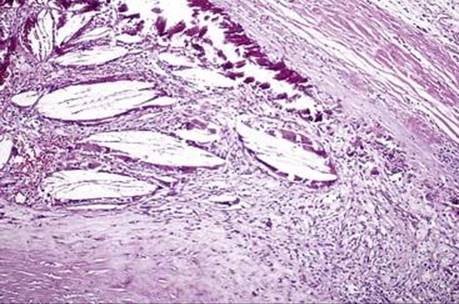
Fig 4 Atherosclerosis. Higher-power view shows prominent calcification and cholesterol debris next to mild chronic inflammation and macrophages.
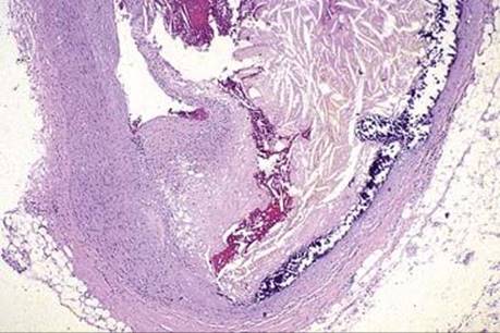
Fig 5 Atherosclerosis. Complicated atherosclerosis with hemorrhage into the plaque after disruption of fibrous cap. Note that the lumen is now severely compromised.