
Definition
• An aortic dissection is a separation of the layers of the blood vessel wall due to hematoma formation by extravasation of blood into the wall
Pathogenesis
• In older adults the most common cause is weakness of the blood vessel wall due to atherosclerosis and hypertension
• In younger adults the most common cause is idiopathic medial degeneration
• Marfan syndrome is a rare cause
Clinical features
• Severe ripping chest pain radiating to the back; can rupture into pericardial sac, resulting in tamponade
• Without treatment 75% to 90% of patients with dissection will die rapidly. With treatment the mortality rate is less than 20%
• Treatment consists of surgical repair and control of hypertension
Pathology
Gross
• Longitudinal tear in wall
• Hematoma within wall of aorta
• False lumen creates a “double-barreled” aorta
Histology
• A second lumen created by the hematoma may be seen on cross-section
• Medial degeneration is commonly seen
• Focal medial necrosis may be present with preserved elastic fibers

Fig 1 Aortic dissection. Gross image of an aortic dissection with a false lumen spanning almost the entire length of the aorta, which has been opened. Note that the endothelial surface of the true lumen is smooth and free of atherosclerosis.
Immunopathology/special stains
• Elastic stain highlights medial degeneration
• Trichrome stain shows focal necrosis of muscle in media

Fig 2 Aortic dissection. Close-up view showing extension of dissection into the abdominal aorta.

Fig 3 Aortic dissection. Gross image of aortic dissection starting just above the aortic valve creating a second lumen. The aorta has been cross-sectioned just above the aortic valve, and the photograph is taken looking toward the valve.
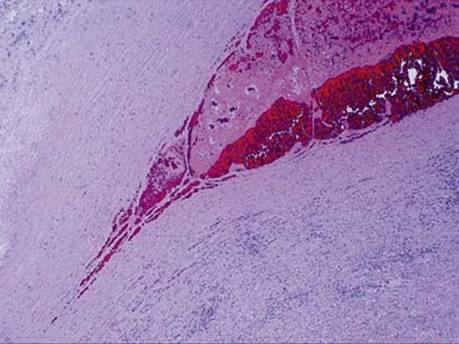
Fig 4 Aortic dissection. Microscopic view of hematoma dissecting through aortic wall.

Fig 5 Aortic dissection. Low-power view showing myxoid degeneration of the media.

Fig 6 Aortic dissection. High-power view with myxoid material and blood within media.

Fig 7 Aortic dissection. High-power view of media in a 40-year-old patient with dissection. The muscle fibers in the pale area are lost; only necrotic elastic fibers are seen.

Fig 8 Aortic dissection. Elastic stain of same area shown in Fig 7 with collapse of elastic fibers onto each other within area of muscle necrosis.

Fig 9 Aortic dissection. Atherosclerotic changes with cholesterol clefts in a 72-year-old patient with aortic dissection.