
Definition
• An aortic aneurysm is a focal abnormal dilation of the aorta to 50% greater than normal diameter
Pathogenesis
• Elastic and collagen breakdown due to an imbalance of metalloproteinases and metalloproteinase inhibitors
• Medial degeneration
• Collagen vascular disease
• Marfan syndrome
• Necrotizing aortitis
• Rarely, caused by infection with Treponema pallidum
• In contrast to abdominal aortic aneurysms, thoracic aortic aneurysms are less likely to be related to atherosclerotic disease, although in older adults it is commonly present
Clinical features
Epidemiology
• Thoracic aneurysms account for less than 10% of all aortic aneurysms
Presentation
• May be asymptomatic or cause chest and or back pain
Prognosis and treatment
• Treated surgically
• If untreated, patients live for an average of 6 to 8 months after the onset of symptoms
Pathology
Gross
• Focal dilation of the aorta
• When due to syphilis, classically occurs in the ascending aorta, which will have a “tree bark” appearance
Histology
• Cystic medial degeneration (cystic spaces filled with myxoid material)
• Atherosclerosis: cholesterol clefts
• Fragmented elastic fibers
• Necrotizing aortitis: focal medial necrosis with mild chronic inflammation and occasional giant cells
• Lymphoplasmacytic infiltrate in adventitia are common
• Luetic aneurysms caused by syphilis classically show obliterative endarteritis of the vasa vasorum (lymphoplasmacytic infiltrate around the small blood vessels within the aortic wall with obliteration of their lumens) and medial necrosis
Immunopathology/special stains
• Elastic stain shows fragmented elastic fibers as opposed to the normal continuous fibers

Fig 1 Thoracic aortic aneurysm. This 72-year-old woman had extensive atherosclerosis leading to thoracoabdominal aneurysms seen here grossly.

Fig 2 Thoracic aortic aneurysm. Rupture of the thoracic aneurysm leading to compression of the esophagus (same patient as in Fig 1). Probe shows the site of rupture.

Fig 3 Thoracic aortic aneurysm. High-power view showing a lymphoplasmacytic infiltrate in the adventitia.

Fig 4 Thoracic aortic aneurysm. Elastic stain reveals fragmented elastic fibers in the aortic wall.
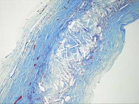
Fig 5 Thoracic aortic aneurysm. Trichrome stain of aneurysm wall with atherosclerosis.

Fig 6 Thoracic aortic aneurysm. Idiopathic necrotizing aortitis: low power shows focal necrosis of media in this aortic aneurysm from a 36-year-old woman.
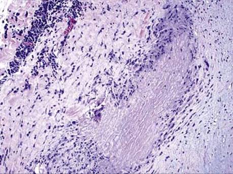
Fig 7 Thoracic aortic aneurysm. Idiopathic necrotizing aortitis: high power shows focal necrosis surrounded by mononuclear cells and a multinucleated giant cell. Adjacent adventitia has a lymphoplasmacytic infiltrate, which is commonly seen in any aneurysm.

Fig 8 Thoracic aortic aneurysm. Idiopathic necrotizing aortitis: other areas of the media show nonspecific degeneration.