
Definition
• Clinical syndrome occurring predominantly in patients with chronic asthma or, less often, bronchiectasis who develop hypersensitivity to Aspergillus fumigatus
Clinical features
Presentation
• Individual with asthma develops peripheral blood eosinophilia, transient pulmonary opacity, elevated total serum IgE, immediate cutaneous reaction to Aspergillus, elevated serum IgG and IgE to Aspergillus, as well as central bronchiectasis
• May have positive sputum culture for aspergillosis
Prognosis and treatment
• Corticosteroid is the main treatment. Itraconazole may play a role in patients with resistance to corticosteroids
• Control the underlying asthmatic disease
Pathology
Histology
• “Allergic mucin” is considered the hallmark of the disease. It is composed of abundant eosinophilic mucin, mixed with eosinophils, eosinophil cytoplasmic debris, occasional Charcot-Leyden crystals as well as calcium oxalate crystals
• Rare fungal hyphae are present in the mucin but do not involve the lung parenchyma or vessels
• Various combinations of asthmatic changes, bronchocentric granulomatosis, and eosinophilic pneumonia are commonly seen
Immunopathology/special stains
• GMS stain highlights the fungal hyphae in most cases, but these are sometimes not identified
Main differential diagnoses
• Eosinophilic pneumonia: eosinophilic inflammation of lung and airways
• Hypersensitivity pneumonia: chronic inflammatory interstitial pneumonia accompanied by poorly formed granulomas with interspersed eosinophils and bronchiolitis
• Invasive fungal pneumonia: granulomatous inflammation with fungal hyphae infiltrating the lung parenchyma and vascular wall
• Churg-Strauss syndrome: combination of eosinophilic pneumonia, asthmatic bronchitis, granulomatous inflammation (allergic granulomas), eosinophilic vasculitis as well as eosinophilic abscesses
• Wegener granulomatosis: systemic disease usually manifested by upper respiratory tract and lung involvement along with glomerulonephritis; associated with increased serum c-ANCA; geographically necrotizing granulomatous inflammation with prominent parenchymal necrosis and vasculitis

Fig 1 Allergic bronchopulmonary aspergillosis. At low power, layers of mucin and inflammatory cells are seen in this plug removed from a 10-year-old with asthma.
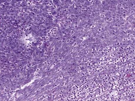
Fig 2 Allergic bronchopulmonary aspergillosis. Depending on the preservation of cells and staining, eosinophils may be difficult to identify as in this figure; binucleation is helpful when the granules are pale.

Fig 3 Allergic bronchopulmonary aspergillosis. High power shows the characteristic “allergic” mucin, which is pale blue and intermixed with numerous eosinophils.

Fig 4 Allergic bronchopulmonary aspergillosis. Charcot-Leyden crystals are seen in this high power of allergic mucin.

Fig 5 Allergic bronchopulmonary aspergillosis. GMS stain will often stain parts of the mucin as seen here on the left. Fragmented fungal hyphae are present in the middle.
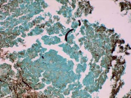
Fig 6 Allergic bronchopulmonary aspergillosis. GMS stain: overstained hyphae can be difficult to identify as seen here.
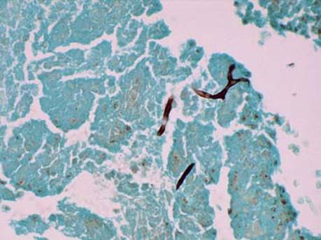
Fig 7 Allergic bronchopulmonary aspergillosis. GMS stain: branching helps to confirm that these black structures are indeed fungal hyphae.