
Definition
• Acute inflammation of bronchioles with variable epithelial sloughing
Clinical features
Epidemiology
• Most often associated with infections, especially in infants and children, toxic fume or gas inhalation, chemotherapy, or acute aspiration
Presentation
• Children with viral infections present with tachypnea, wheezing, and prolonged expirations
• Adults tend to have few specific symptoms
Prognosis and treatment
• Most patients fully recover, especially from infectious causes
• Patients with an idiopathic cause may respond to antibiotics and immunosuppressive therapy but often progress to decreased lung function
Pathology
Histology
• Extension of acute inflammation into the walls of bronchioles and intraluminal collections of neutrophils as well as mucus
• Frequently associated with chronic bronchiolitis
Immunopathology/special stains
• Not contributory
Main differential diagnoses
• Bronchopneumonia: acute inflammation extending from the airways into the adjacent alveoli
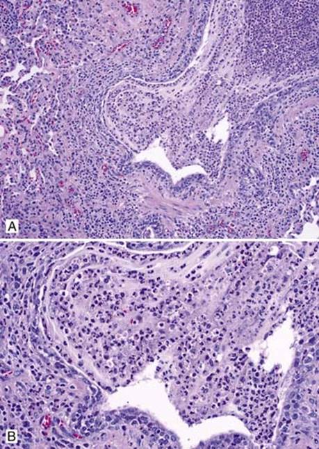
Fig 1 Acute bronchiolitis. A, Inflammatory cells infiltrate predominantly the bronchiolar wall; mucostasis with abundant neutrophils in the lumen. B, High magnification reveals that mixed inflammatory cells, with neutrophils predominant, expand the subepithelial space; the bronchiolar epithelial cells are attenuated and focally detached; accumulated mucus with mixed numerous neutrophils.
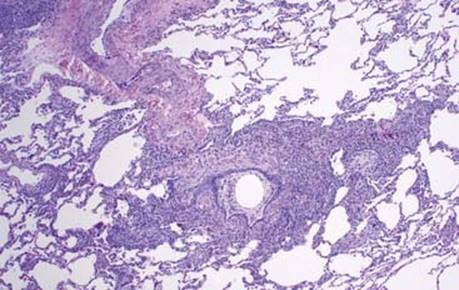
Fig 2 Acute bronchiolitis. Mixed acute and chronic inflammation with mucostasis is seen here. The inflammation extends into peribronchiolar connective tissue, but most of the adjacent alveolar spaces are open.
Chronic Bronchiolitis
Definition
• Chronic inflammation of the small airways characterized by mixed chronic inflammatory cells within and surrounding the bronchiolar walls
Clinical features
• Nonspecific histological finding, usually associated with a wide variety of lung diseases, including infection, autoimmune diseases, asthma, drugs, radiotherapy, chemotherapy, and transplantation
• Infrequently encountered as the only abnormality on a lung biopsy specimen; its clinical significance is unclear
Prognosis and treatment
• Varies depending on underlying diseases
Pathology
Histology
• Chronic inflammatory cells infiltrating the bronchiolar wall and surrounding peribronchiolar connective tissue
• May see accompanying goblet cell hyperplasia, smooth muscle hyperplasia, and mild luminal narrowing
• Sometimes, prominent accumulation of foamy macrophages infiltrate the bronchiolar walls and accumulate in the lumens
Immunopathology/special stains
• Not contributory
Main differential diagnosis
• Follicular bronchiolitis: chronic inflammation of the small airways with formation of lymphoid follicles containing germinal centers

Fig 1 Chronic bronchiolitis. The mixed chronic inflammatory cells infiltrate the submucosa and are present through the smooth muscle layer of the small airway. The bronchiolar epithelium and adjacent alveoli are unremarkable.

Fig 2 Chronic bronchiolitis. The airway is surrounded by lymphocytic infiltrate and variable distortion with focal bronchiolar epithelial sloughing and luminal mucus mixed with neutrophils. Atelectasis is present in the adjacent alveoli.

Fig 3 Chronic bronchiolitis. Mild fibrosis is present in the submucosa accompanied by minimal luminal narrowing. Chronic inflammation is less prominent.
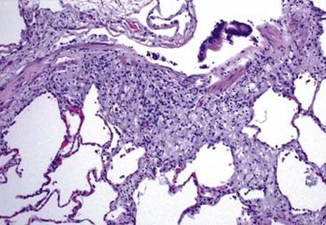
Fig 4 Chronic bronchiolitis. In this case, predominant foamy macrophages infiltrate the bronchiolar wall and extend into the adjacent lung. There are very few inflammatory cells. The bronchiolar epithelial cells are largely sloughed.