
Definition
• Acute necrotizing vasculitis involving arterioles, capillaries, and venules (also called microscopic polyarteritis, hypersensitivity vasculitis, or leukocytoclastic vasculitis)
Clinical features
Epidemiology
• Incidence increases with age
• No gender predilection
• Associated with connective tissue diseases, certain drugs, tumors, Henoch-Schönlein purpura, essential mixed cryoglobulinemia
• Postulated to be a hypersensitivity reaction to drugs, tumor antigens, and organisms, among others
Presentation
• Involves smaller vessels than polyarteritis nodosa
• Signs and symptoms depend on organs involved: hemoptysis (lung), myalgia (muscle), abdominal pain (gastrointestinal tract), hematuria or proteinuria (kidneys)
• Cutaneous involvement is seen as “palpable purpura” of the skin or rash
• Isolated cutaneous involvement is termed cutaneous leukocytoclastic vasculitis
• No hypertension (in contrast to polyarteritis nodosa)
• About 70% of cases are associated with p-antineutrophil cytoplasmic antibody (ANCA)
• Diagnosis requires fulfilling three or more of the following diagnostic criteria (from Calabrese LH, Michel BA, Bloch DA, et al: The American College of Rheumatology 1990 criteria for the classification of hypersensitivity vasculitis, Arthritis Rheum 33:1108-1113, 1990):
• Age older than 16 years
• Medication at onset of disease (presumed precipitating factor)
• Palpable purpura
• Maculopapular rash
• Biopsy demonstrating leukocytoclastic vasculitis
Prognosis and treatment
• Most cases respond to removal of precipitating antigenic stimulus
• Some cases with widespread vasculitis are more aggressive
Pathology
Histology
• Segmental fibrinoid necrosis of vascular media
• Neutrophils infiltrate vascular wall; neutrophilic debris (fragmented and disintegrated neutrophils) is also present
• Vasculitic lesions are the same age
• No granulomas (in contrast to Wegener granulomatosis)
Immunopathology/special stains
• Not contributory
Main differential diagnoses
• Polyarteritis nodosa: vasculitic lesions are of different ages and involve somewhat larger vessels; patients have hypertension
• Churg-Strauss syndrome: granulomas and eosinophils; associated with bronchial asthma and peripheral eosinophilia
• Wegener granulomatosis: associated with c-ANCA; necrotizing granulomas

Fig 1 Microscopic polyangiitis. Ulceration of rectal mucosa due to involvement by microscopic polyangiitis.

Fig 2 Microscopic polyangiitis. Neutrophils are present within the walls of involved vessels (A and B).

Fig 3 Microscopic polyangiitis. The infiltrating neutrophils undergo “leukocytoclasia,” or disintegration and fragmentation.
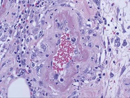
Fig 4 Microscopic polyangiitis. Not all vessels with fibrinoid necrosis have neutrophils.

Fig 5 Microscopic polyangiitis. Dermal involvement by leukocytoclastic vasculitis shows small vessel involvement with extravasated neutrophils and red blood cells (A and B).