
Definition
• Idiopathic, segmental, acute, and chronic thrombosing vasculitis involving small and medium-sized peripheral arteries and veins (also called Buerger disease)
Clinical features
Epidemiology
• Rare disease (8 to 12.6 cases per 100,000) affecting predominantly men younger than 35 years
• Strong association with smoking
• Associated with HLA-A9 and HLA-B5 haplotypes
• More common in Israel, Japan, and India; less common in the United States and Europe
Presentation
• Usually involves the tibial and radial arteries
• May spread to adjacent veins and nerves
• Raynaud phenomenon (cold sensitivity), severe pain in affected extremity, intermittent claudication, superficial phlebitis
• Most recent set of diagnostic criteria (from Olin JW: Thromboangiitis obliterans (Buerger’s disease), N Engl J Med 343:864-869, 2000):
• Age younger than 45 years
• Current or recent history of tobacco use
• Evidence of distal extremity ischemia (e.g., claudication, ulceration, gangrenous necrosis, positive vascular testing)
• Exclusion of other causes of peripheral vascular disease, such as diabetes mellitus, hypercoagulable states, or autoimmune diseases
• Exclusion of proximal source of emboli by echocardiography or arteriography
• Confirmatory arteriogram in involved and uninvolved extremities
Prognosis and treatment
• May lead to gangrenous necrosis of affected limb, which requires amputation
• Smoking cessation is the only treatment that prevents progression of disease
Pathology
Histology
• Early lesion: thrombosis and transmural acute and chronic inflammatory infiltration of vessel wall
• Thrombus contains central core of neutrophils (microabscess) surrounded by granulomatous inflammation
• Later lesion resembles organized thrombus with stenosis or complete luminal occlusion
• Affected arteries, veins, and nerves eventually encased in fibrosis
Immunopathology/special stains
• Not contributory
Main differential diagnoses
• Polyarteritis nodosa: no association with smoking; presents with hypertension; vasculitic lesions of different ages
• Microscopic polyangiitis: smaller vessels involved; associated with p-ANCA; leukocytoclastic vasculitis rather than thrombosis; cutaneous manifestations (palpable purpura and maculopapular rash)
• Thromboembolic disease: proximal source of embolus; atherosclerosis involving larger vessels
• Peripheral vascular disease: atherosclerotic lesions, which involve larger arteries than those in thromboangiitis obliterans
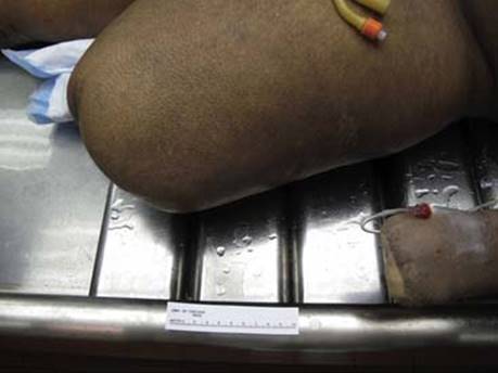
Fig 1 Thromboangiitis obliterans. Postmortem gross photograph of patient with gangrenous necrosis secondary to Buerger disease after triple amputation, showing the left upper and lower extremities’ stumps.
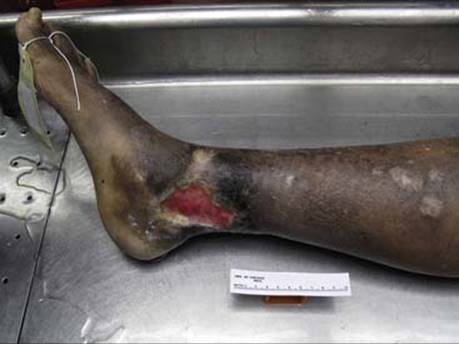
Fig 2 Thromboangiitis obliterans. Gross photograph of ischemic ulcer in the residual extremity of patient in Fig 1.
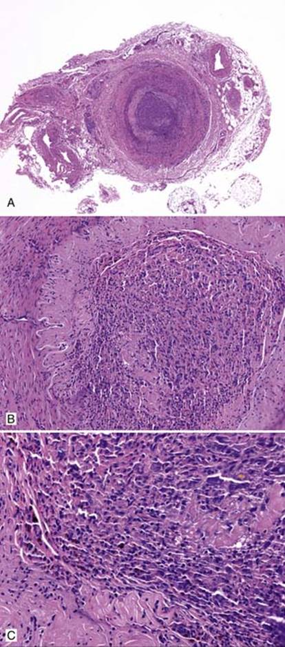
Fig 3 Thromboangiitis obliterans. Low (A), intermediate (B), and high (C) powers of artery occluded by chronic inflammatory infiltrate including eosinophils.
(Courtesy of Dr. Haresh Mani, Hershey, Pa.)
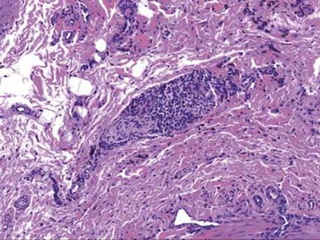
Fig 4 Thromboangiitis obliterans. Adjacent nerve is also involved by chronic inflammation.
(Courtesy of Dr. Haresh Mani, Hershey, Pa.)