
Definition
• Presence of lymphoid follicles next to and confined to the walls of the airways and associated peribronchial tissues
Clinical features
Epidemiology
• May be associated with collagen vascular diseases (e.g., rheumatoid arthritis), immunodeficiency syndromes (e.g., IgA deficiency, congenital variable immunodeficiency, AIDS), bronchiectasis, obstructive pneumonias, chronic infection, and hypersensitivity syndromes
Presentation
• Varies depending on the underlying disease
Prognosis and treatment
• Varies depending on the underlying disease
Pathology
Histology
• Lymphoid follicles containing reactive germinal centers are present in the walls of bronchioles and occasionally pleura, especially in patients with rheumatoid arthritis
• There may be postobstructive changes such as mucostasis and organizing pneumonia
• Absence of significant lymphoid infiltrate in the adjacent lung parenchyma
Immunopathology/special stains
• Not contributory
Main differential diagnoses
• Lymphoid interstitial pneumonia: diffuse interstitial infiltrate of mature lymphocytes and admixture of chronic inflammatory cells; associated with infections (HIV, Epstein-Barr virus, hepatitis viruses) and autoimmune diseases (Sjögren); the spectrum of radiographic changes varies from bilateral streaky densities to reticulonodular opacities
• Nodular lymphoid hyperplasia: considered to be late stage of a healing inflammatory process; nodular lymphoid proliferation associated with fibrosis; masslike lesion on radiographic image
• Lymphomas: clonal expansion of neoplastic lymphoid cells with abnormal immunophenotype and cytogenetic or molecular studies; commonly encountered lymphomas include chronic lymphocytic lymphoma, follicular center cell lymphoma, mantle cell lymphoma, MALT lymphoma, Hodgkin lymphoma
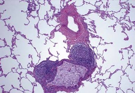
Fig 1 Follicular bronchiolitis. Lymphoid follicles confined to the wall of airway; no significant lymphoid involvement in the adjacent lung parenchyma. The airway is dilated and has mucostasis.

Fig 2 Follicular bronchiolitis. Follicular bronchiolitis with adjacent lung parenchyma showing foci of organizing pneumonia, probably after obstruction.
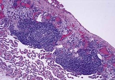
Fig 3 Follicular bronchiolitis. Follicular pleuritis is seen in this patient with rheumatoid arthritis.