
Definition
• Destruction of acinar walls resulting in permanent enlargement of airspaces that are distal to terminal bronchioles; usually no significant fibrosis
• Subclassified into three types according to the portion of affected pulmonary acini: proximal acinar (centrilobular) emphysema, panacinar emphysema, and distal acinar emphysema
Clinical features
Epidemiology
• Tobacco smokers most often have proximal acinar emphysema
• Patients with α1-antitrypsin (A1AT) deficiency usually develop panacinar emphysema
• Patients with spontaneous pneumothorax have distal acinar or irregular emphysema
• Other risk factors include marijuana abuse and dust and chemical exposure
Presentation
• Progressive dyspnea
• Decreased breath sounds, crackles at the bases, distant heart sounds, and depressed diaphragm
• Radiographic images: evidence of overinflation characterized by increased radiolucency of the lungs, flat diaphragm, decreased heart shadow
• Pulmonary function tests: airflow obstruction
Prognosis and treatment
• Stop smoking and avoid adverse stimulators to halt further lung damage
• Patient with A1AT deficiency: goal of treatment is to increase A1AT level in serum and lung interstitium; current major approach is intravenous augmentation therapy by infusion of pooled human A1AT
• Treated symptomatically with anticholinergics, bronchodilators, corticosteroids as well as supplemental oxygen
• At the late stage of the disease, the damage becomes irreversible; patient may need lung volume reduction surgery or a lung transplant
Pathology
Gross
• Terminal airways are dilated and form many cavities
Histology
• Microscopically, there is destruction of acinar walls and dilatation of the airspaces
• Bronchial metaplasia is often present
• Usually not associated with significant inflammation or fibrosis
• Panacinar emphysema: the acinus is uniformly involved; the lower lobes are more predominantly involved
• Proximal acinar emphysema: proximal portion of the acinus around respiratory bronchiole is predominantly involved; upper lobes are more severely affected
• Distal acinar emphysema: the distal acinus, along the interlobular septa and close to pleura, is affected; may form blebs and bullae; spontaneous pneumothorax
Immunopathology/special stains
• Not contributory
Main differential diagnosis
• Congenital lobar emphysema: occurs in infants, congenital malformation of the lung characterized by hyperinflation of one or more pulmonary lobes
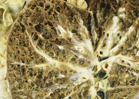
Fig 1 Emphysema. Gross photograph of cut surface of formalin-inflated lung shows relatively uniform enlarged acini in this patient with A1AT deficiency.
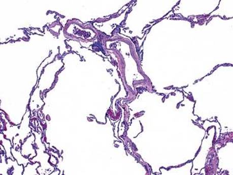
Fig 2 Emphysema. Alveolar spaces are markedly enlarged while the alveolar walls are of normal thickness (i.e., without fibrosis).
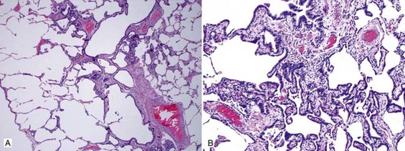
Fig 3 Emphysema. Emphysema and bronchial epithelial metaplasia (occurring in a smoker): some of the alveolar walls are lined by metaplastic bronchial epithelium; low (A) and high (B) powers. Note focal interstitial fibrosis.
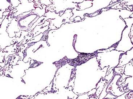
Fig 4 Emphysema. Anthracotic pigment is often present in smokers with emphysema, as seen here.
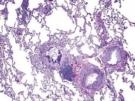
Fig 5 Emphysema. Pulmonary hypertensive changes (intimal fibrosis and muscle hyperplasia) are present in this patient with emphysema.