
Definition
• An abnormal accumulation of fluid in pulmonary interstitium and alveoli
Pathogenesis
• Increased vascular permeability due to a spectrum of endothelial and epithelial injuries of the lung leading to one or more of the following:
• Increased capillary hydrostatic pressure
• Increased capillary permeability
• Decreased plasma oncotic pressure
• Lymphatic obstruction
Clinical features
Epidemiology
• Incidence varies with inciting cause; left-sided heart failure is the main cause
• Other reasons include lung injury, pneumonia, toxins, medications, and living at high altitude
Presentation
• Shortness of breath, anxiety, and a feeling of drowning
• In patients with long-standing pulmonary edema there may be dyspnea on exertion, orthopnea, paroxysmal nocturnal dyspnea, or coughing
• Radiological features include butterfly pattern of lung infiltrates, pleural effusion(s), Kerley B lines, and loss in radiological sharpness of the vasculature
Prognosis and treatment
• Pulmonary edema can be fatal; therapy and outcome depend on the underlying etiology
• 20% mortality in intensive care unit patients
• Prompt treatment of pulmonary edema along with treatment of the underlying cause improves survival
Pathology
• Gross appearance: heavy lungs, frothy exudates, and dark blue to red cut surface. In long-standing pulmonary edema the cut surface may be firm and brown
• Microscopic appearance
• Pale pink homogenous fluid in alveolar spaces
• Congestion
• Leakage of some red blood cells into airspaces
• In patients with long-standing pulmonary edema, hemosiderin-laden macrophages are present and there may be alveolar septal thickening and fibrosis
Main differential diagnoses
• Diffuse alveolar damage (DAD): presence of hyaline membranes
• Alveolar proteinosis: accumulation of granular proteinaceous material in alveoli
• Fibrin exudates: dark pink strands of fibrin can be seen
• Pneumocystis jiroveci pneumonia

Fig 1 Pulmonary edema. H&E stain of lung section showing alveoli filled with pink proteinaceous material in a patient with heart failure. Note congested vessels in alveolar septa.

Fig 2 Pulmonary edema. Pulmonary edema is often not uniform: some alveoli are completely filled and others only partially, as seen in this figure.
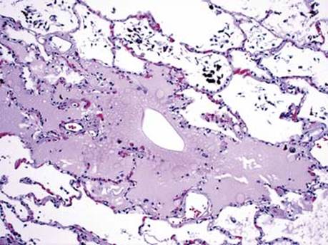
Fig 3 Pulmonary edema. Prussian blue stain highlights hemosiderin-laden macrophages in this patient with congestive heart failure who also has intraalveolar edema.

Fig 4 Pulmonary edema. Ventilator therapy pushes the pale edema fluid to the walls of the alveoli, which suggests the differential diagnosis of DAD.

Fig 5 Pulmonary edema. Hyaline membranes in DAD are bright pink (eosinophilic).