
Definition
• An autoimmune disease characterized by T-lymphocyte infiltration of salivary glands with atrophy and dryness of mucous membranes; frequently involves the lungs
Clinical features
Epidemiology
• Half of cases occur as primary SS, whereas half occur as SS associated with other connective tissue diseases
• Lung involvement is more common in women with SS than in men
Presentation
• Dry cough, dyspnea, and recurrent respiratory infections in a patient with xerostomia, xerophthalmia, and arthritis
• Positive serological tests for ANA, RF, anti-SSA, and anti-SSB
Prognosis and treatment
• Good prognosis with corticosteroids and other immunosuppressant therapy
• Usual interstitial pneumonia (UIP) pattern may have worse prognosis
Pathology
Histology
• Most common finding is of nonspecific interstitial pneumonia (NSIP), characterized by a bronchiolocentric uniform thickening of alveolar septa by lymphocytes and/or fibrosis and associated type II pneumocytic hyperplasia
• Mucous glands of the large airways are atrophic and have lymphocytic infiltrates
• Small, nonnecrotizing interstitial granulomas and multinucleated giant cells may be seen
• Lymphocytic infiltrates may obscure underlying lung architecture and may be due to benign entities, such as lymphoid interstitial pneumonia or follicular bronchiolitis, or to malignant entities such as lymphomatoid granulomatosis or lymphoma
• Secondary pulmonary infection in the presence of significant lymphocytic infiltrates due to decreased mucous production and abnormal mucociliary clearance from the large airways
• Occasionally bronchiectasis, pulmonary cysts, and bullae may be present
• UIP pattern of geographical and temporal heterogeneity of interstitial fibrosis with microscopic honeycombing is seen occasionally
• Other infrequent findings include amyloidosis, pulmonary hypertensive changes, pulmonary hemorrhage, and pleuritis
Immunopathology/special stains
• Not contributory
Main differential diagnoses
• Other causes of NSIP and UIP
• Other lung diseases with granulomas and multinucleated giant cells, such as hypersensitivity pneumonitis, infection, and aspiration
• Primary lymphoproliferative lesions or lymphomas of the lung

Fig 1 Sjögren syndrome. NSIP and pleural fibrosis are seen in this wedge biopsy from a patient with SS.

Fig 2 Sjögren syndrome. Extensive interstitial inflammation, as seen here, is often seen in lung disease associated with SS.

Fig 3 Sjögren syndrome. UIP pattern with pleural fibrosis and chronic inflammation with lymphoid aggregates and germinal centers is seen in this case of SS-associated lung disease.
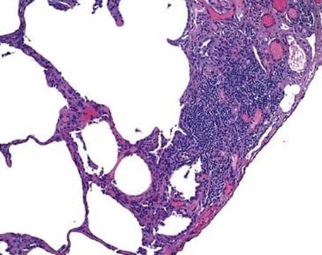
Fig 4 Sjögren syndrome. Lymphoid aggregates can be seen abutting the pleura.
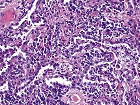
Fig 5 Sjögren syndrome. Type II pneumocyte hyperplasia and extensive mixed inflammatory infiltrate of the interstitium is seen at high power.