
Definition
• Interstitial lung disease arising in patients with a gastrointestinal chronic inflammatory condition manifesting as ulcerative colitis or as Crohn disease
Clinical features
Epidemiology
• Up to half of IBD patients may have respiratory tract involvement
• Respiratory symptoms are more common in ulcerative colitis than in Crohn disease
• Women are more often affected than men; age of onset is variable
• Pulmonary involvement typically occurs in patients known to have IBD but infrequently may be the presenting condition
• The degree of inflammatory changes in the lungs tends to correlate with that in the gastrointestinal tract
Presentation
• Airway inflammation: coughing, wheezing, obstruction, abnormal pulmonary function tests
• Pulmonary parenchymal disease: dyspnea, coughing, fever, abnormal pulmonary function tests, basilar lung opacities or infiltrates on chest CT
Prognosis and treatment
• High-dose corticosteroids
• Prognosis varies with type and extent of pulmonary involvement
Pathology
Histology
• Airway inflammation: tracheitis, bronchitis, bronchiectasis, acute or chronic bronchiolitis, bronchopneumonia-like pattern, lung abscess
• Parenchymal disease: interstitial lung disease (most often nonspecific interstitial pneumonia), organizing pneumonia, eosinophilic pneumonia
• Nonnecrotizing granulomas may be present in Crohn disease
• Pulmonary thromboemboli
• Patients with IBD-associated pyoderma gangrenosum of the skin may rarely have similar necrobiotic nodules with acute and chronic inflammatory cells in the lungs
Immunopathology/special stains
• Not contributory
Main differential diagnoses
• Sulfasalazine or mesalamine (used in IBD) therapy-related pneumonitis: frequently associated with peripheral blood eosinophilia; more alveolar than airway involvement
• Infection
• Other causes of interstitial lung disease
• Hypersensitivity pneumonitis
• Sarcoidosis
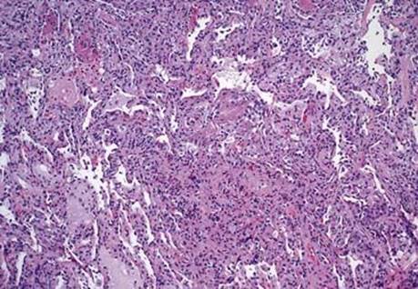
Fig 1 Inflammatory bowel disease–associated interstitial lung disease. Eosinophilic pneumonia is present in this transbronchial biopsy from a patient with Crohn disease.
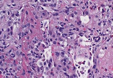
Fig 2 Inflammatory bowel disease–associated interstitial lung disease. At higher power, several eosinophils and an eosinophilic alveolar exudate are seen.
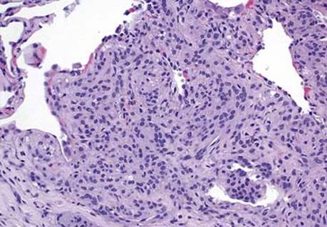
Fig 3 Inflammatory bowel disease–associated interstitial lung disease. The presence of granulomas can be a clue to Crohn disease–associated lung disease; however, a meningothelial-like nodule, as seen here, should not be mistaken for a granuloma.