
Definition
• Pulmonary interstitial fibrosis caused by inhalation of asbestos fibers
Pathogenesis
• Due to substantial and significant exposure to asbestos fibers
• Different forms of asbestos:
• Serpentine/chrysotile:
– Curly and flexible
– More commonly seen in industry but less pathogenic
– Usually caught in upper respiratory tract and removed with mucus by coughing
• Amphibole:
– Straight and stiff
– More pathogenic
– Usually goes deep in lung; reaches small airways
• Both forms are fibrogenic; macrophages at the small airways ingest fibers and release chemotactic factors and fibrogenic mediators, causing peribronchiolar and interstitial fibrosis
• Asbestos exposure also increases the risk of malignancy (lung cancer and mesothelioma)
• Asbestos fibers may also absorb toxic chemicals from tobacco smoke; therefore, asbestos exposure increases the risk of lung cancer significantly (10 times higher risk in smokers than in those with asbestos exposure alone)
Clinical features
Epidemiology
• Occurs in a variety of occupational settings:
• Asbestos mining and milling
• Asbestos products manufacturing: insulator, shipyard worker, construction worker, and others
• Household contacts of asbestos workers
• Long latent period, usually develops 10 years or more after exposure
Presentation
• Initially shortness of breath on exertion, later dyspnea even at rest; may progress to heart failure with hypoxia and cor pulmonale
• Restrictive changes on pulmonary function tests
• Disease starts from lower lobes and subpleurally; later extends to middle and upper lobes
Prognosis and treatment
• Treatment: stop asbestos exposure and stop smoking; relieve symptoms and prevent complications
• Disease is irreversible; prognosis depends on the amount of lung fibrosis
Pathology
Histology
• Initially, peribronchiolar fibrosis with asbestos bodies
• Later, diffuse interstitial fibrosis with honeycombing, mucostasis, and type II pneumocytic hyperplasia with eosinophilic cytoplasmic hyaline; rarely, fibroblastic foci may be seen, mimicking usual interstitial pneumonia (UIP)
• Some have prominent intraalveolar macrophages, which resemble desquamative interstitial pneumonia
• Asbestos body is the hallmark finding: a beaded or dumbbell-shaped rod with a central clear core and golden brown coating; it may be straight or curved
• Asbestos bodies can be found in peribronchiolar macrophages, alveolar spaces or septum, or even within thoracic lymph nodes
• Ferruginous body is a general term for an inorganic particle coated with iron; asbestos body is a ferruginous body that contains asbestos fibers coated with iron-containing proteinaceous material
Immunopathology/special stains
• Iron stain: helps identify asbestos bodies by staining the iron coating blue
• Electron microscopy: facilitates the characterization of asbestos fibers
Main differential diagnoses
• UIP: lacks asbestos bodies
• Other forms of diffuse pulmonary fibrosis
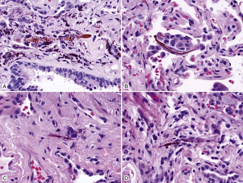
Fig 1 Asbestosis. Asbestos bodies can be seen in peribronchiolar macrophages (A), intraalveolar space (B), and alveolar septum or interstitium (C and D). Note beaded, rod-like structures with a central core and variable length.
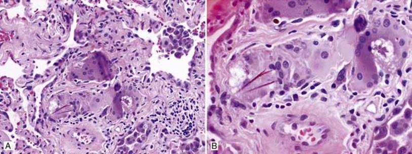
Fig 2 Asbestosis. Multinucleated giant cells containing asbestos bodies and asteroid bodies: low (A) and medium (B) powers.
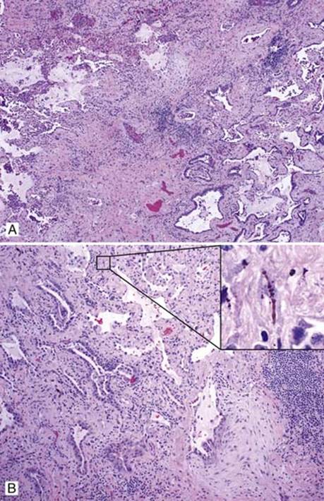
Fig 3 Asbestosis. Asbestosis in a UIP pattern with interstitial fibrosis, microscopic honeycombing, and fibroblastic focus: A, low power; B, medium power showing fibroblastic focus; inset, demonstrating amplified asbestos fiber.
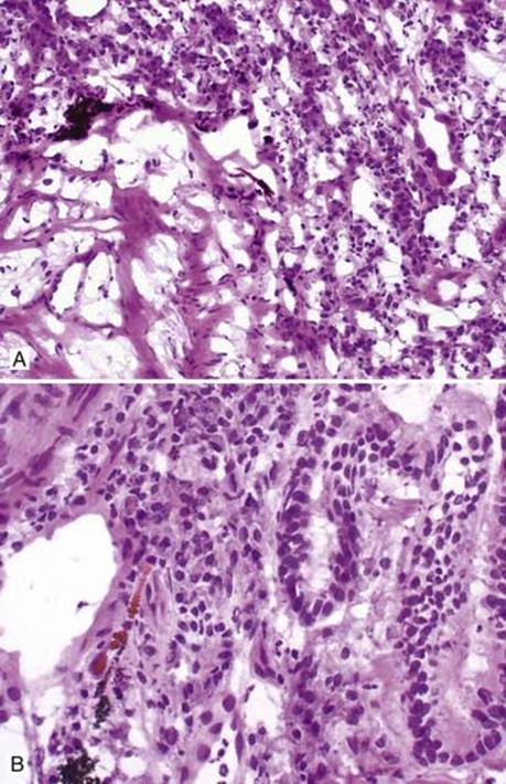
Fig 4 Asbestosis. Asbestos bodies can also be identified on frozen section: low (A) and high (B) powers. This frozen section was done for diagnosis of adenocarcinoma, which is shown in B.