
Definition
• A progressive parenchymal lung disease resulting from coal dust inhalation
Pathogenesis
• Coal is a fossil fuel, and the most significant source of coal dust exposure is coal mining. Although the major component—carbon—is relatively inert, coal mine dust also contains hydrogen, oxygen, nitrogen, trace metals, inorganic minerals, and crystalline silica, many of which may be cytotoxic
• Anthracite coal mining has been associated with higher rates of pneumoconiosis than that found in bituminous miners, probably because the former contains more surface free radicals as well as a greater content of crystalline silica
• Cumulative exposure to dust determines progression to progressive massive fibrosis (PMF). Other factors that may impact progression to PMF include high silica content of the mine dust, infection with Mycobacterium tuberculosis, and immunological factors
Clinical features
Epidemiology
• The incidence and rate of CWP progression is related to the amount of respirable coal dust to which miners were exposed during their working lifetime
• Approximately 200,000 workers are employed in the coal mining industry in the United States. In a recent study of 145,512 miners, it was found that the prevalence of CWP is increasing in mines of all sizes, while CWP and PMF are much more prevalent among workers from underground mines with fewer than 50 workers (“small mines”)
• A National Institute for Occupational Safety and Health analysis estimated that during the period 1968 to 2006, a total of 22,625 years of potential life lost before age 65 years (YPLL) were attributed to CWP (mean per decedent, 5.7). Disturbingly, annual YPLL from CWP increased from 135 YPLL in 2002 to 169 YPLL in 2006, suggesting a need for strengthening CWP prevention measures
Presentation
• Coal miners typically develop one of two forms of disease patterns—simple CWP or complicated CWP:
• Simple form: chest radiographs in simple CWP show small, round nodular opacities and, occasionally, reticular or reticulonodular opacities (1-5 mm). Calcifications are seen in up to 20% of patients. CT scan features of CWP may be similar to those of silicosis with upper lobe dominant diffusely distributed small nodules in a perilymphatic distribution
• Complicated form: with chronic exposure, the milder form of CWP may become complicated CWP, with enlargement and fibrosis. This form is synonymous with PMF. These patients often have mixed restrictive and obstructive defects on pulmonary function tests and have hypoxemia. On chest radiographs, large opacities may be seen, similar to that in complicated silicosis. Radiologically, PMF may be confused for lung cancer. The distinction between these two may be possible on MR imaging (high signal intensity on T2-weighted images in lung cancer versus low signal intensity in PMF). FDG-PET may show high uptake in fibrotic masses of PMF and is not helpful in making the distinction
• The term black lung is more a legal term than a medical term and is used to include a wide range of lung diseases (including CWP, bronchitis, emphysema, and silicosis) found in association with employment history in coal mines
Prognosis and treatment
• Patients with CWP are at risk for tuberculosis, as are those with silicosis
• Chronic obstructive pulmonary disease is a common association and contributes to mortality
• Workers with a high degree of PMF have a significantly increased mortality rate. They become progressively hypoxemic and may develop cor pulmonale, despite cessation of coal dust exposure. It is estimated that 4% of coal miners die of causes directly related to CWP
Pathology
Histology
• Simple CWP is characterized by the presence of black coal dust macules. These macular lesions are seen around respiratory bronchioles, mostly in the upper lobes of the lung. They range in size from 1 to 6 mm in diameter and are irregular in shape. They are composed of coal dust–laden macrophages with a fine network of reticulin but no fibrosis. Associated centriacinar emphysema (so-called focal emphysema) is characteristic. These lesions are usually asymptomatic. As with silicosis, septal and subpleural deposits (lymphatic distribution) may be present
• When there is significant silica content in the inhaled dust, patients develop palpable nodular lesions, as in silicosis. However, in CWP, these nodules contain abundant black pigment in addition to slate gray silica (“silicoanthracotic nodules”). Based on size, the nodules are classified as micronodules (up to 0.7 cm) or macronodules (0.7 to 2 cm)
• PMF predominantly affects upper and posterior lung zones and is asymmetric in distribution. Gross lesions are at least 2 cm in size and are rubbery black. Cavitation may occur and, as in silicosis, suggests tuberculosis or ischemic necrosis. The necrotic material is black. Unlike in complicated silicosis, the fibrotic mass of CWP consists of haphazardly arranged collagen with abundant intervening pigment, both free and within macrophages. Vascular structures are also often obliterated. The fibrous masses are characteristically nonnodular and have irregular borders with surrounding cicatricial emphysema. Histological overlap, however, may be seen with silicosis, depending on the silica content in the inhaled coal dust. Numerous silicate particles may be seen under polarized light
• Caplan syndrome—this refers to the presence of pulmonary nodules in coal miners with circulating rheumatoid factor. It is also referred to as rheumatoid pneumoconiosis. It has been reported most frequently in Welsh miners. Macroscopically, the nodules range from 0.5 to 5 cm in size and are pale yellow, with eosinophilic granular centers. Microscopically, the centers show necrobiosis, and the periphery of the lesion is composed of concentrically arranged collagen with lymphocytes, plasma cells, and palisaded histiocytes, similar to rheumatoid nodules. It has been described in patients with circulating rheumatoid factor even in the absence of clinical arthritis. It has also been described in those exposed to silica
Immunopathology/special stains
• Not contributory for diagnosis
• AFB stains in cavitary and necrotic lesions to evaluate for tuberculosis
Main differential diagnoses
• Silicosis: nodules are well-defined at the margins and more solid/uniform in appearance on radiology. Calcification develops as a central nodular dot in CWP but tends to be more diffuse in silicosis. Eggshell calcification of hilar nodes is more typical of silicosis. Histologically, silicotic nodules may be a component of both diseases, although CWP also shows abundant black pigment
• Simple anthracosis: much more common than CWP. Black pigment accumulation may be seen in lungs and hilar nodes of cigarette smokers and most adults residing in urban industrialized regions. Occupational history is therefore paramount in the diagnosis of CWP
• Graphite workers’ pneumoconiosis: although graphite miners may have the same pathological changes observed in silicosis and CWP, they also commonly have intraalveolar giant cells. Dust from graphite consists of approximately 50% crystalline carbon or graphite and 25% quartz; the remainder is composed of various silicates
• Carbon electrode makers’ pneumoconiosis: may have pathological changes similar to CWP
• Lung cancer
• Idiopathic pulmonary fibrosis: a usual interstitial pneumonia pattern of fibrosis has been described in PMF; however, there is only minimal to mild anthracosis in idiopathic pulmonary fibrosis
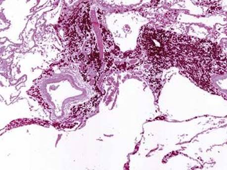
Fig 1 Coal workers’ pneumoconiosis. Dust macules show peribronchiolar and perivascular collection of black dust–laden macrophages accompanied by little collagen fibrosis. Mild centrilobular emphysema is seen.
(Case courtesy of Dr. Dani S. Zander, Hershey, Pa.)

Fig 2 Coal workers’ pneumoconiosis. Large airways may also be affected.
(Case courtesy of Dr. Dani S. Zander, Hershey, Pa.)

Fig 3 Coal workers’ pneumoconiosis. Coalescence of nodules and dense fibrosis is seen in PMF (A). Note the abundant black pigment in the background. A higher-power view shows adjacent cicatricial emphysema (B).
(Case courtesy of Dr. Dani S. Zander, Hershey, Pa.)