
Definition
• Multisystem granulomatous disease of unknown cause with frequent lung involvement
Clinical features
Epidemiology
• Frequently affects young to middle-aged adults
• Higher risk in blacks and females
• Rare in Asians
Presentation
• Majority asymptomatic or with mild, nonspecific pulmonary complaints of dyspnea, coughing
• Abnormal pulmonary function tests such as restrictive defects or low diffusing capacity
• Radiographic abnormalities: bilateral interstitial opacities associated with hilar lymph node enlargement or, less frequently, solitary or multiple nodular densities; localized area of consolidation
Prognosis and treatment
• Variable clinical course
• Slowly progressive disease, although most patients experience remission after treatment
• Some develop extensive pulmonary fibrosis and may require lung transplantation
• Treatment includes immunosuppressants and antifibrotic agents in symptomatic patients
Pathology
Histology
• Classic sarcoidosis
• Nonnecrotizing granulomatous inflammation is distributed primarily along the bronchovascular bundle and lymphatics
• The granulomas predominantly involve airway submucosa and the pulmonary interstitium rather than airspaces; up to 10% of cases have pleural involvement
• Diagnostic tissue is often obtained by transbronchial and endobronchial biopsies
• Accompanied by granulomatous vasculitis characterized by the presence of nonnecrotizing granulomas within the intima and media of blood vessels without causing necrosis of the blood vessel walls
• Granulomas are well circumscribed and composed of tightly clustered epithelioid histiocytes and occasional multinucleated giant cells with few intervening lymphocytes and other inflammatory cells
• Nonspecific cytoplasmic inclusions may be present within giant cells and histiocytes, such as asteroid or Schaumann/conchoid bodies
• Hyalinized fibrous tissue may replace portions of granulomas or even entire granulomas
• Necrotizing sarcoid granulomatosis
• Differs histologically from classic sarcoidosis by the presence of large areas of parenchymal necrosis in the middle of confluent granulomas as well as nonnecrotizing granulomas
• More widespread and prominent granulomatous vasculitis
Immunopathology/special stains
• Epithelioid histiocytes are positive for CD68
• Special stains and tissue culture are needed to rule out acid-fast bacilli and fungal infection
• Serum angiotensin-converting enzyme is elevated in 30% to 80% of patients
Main differential diagnoses
• Infectious diseases: fungi (histoplasmosis, blastomycosis, or coccidioidomycosis) and acid-fast mycobacteria
• Hypersensitivity pneumonia: poorly formed granulomas with eosinophils; no lymph node involvement
• Wegener granulomatosis: geographical dirty blue necrotizing granulomas and serological abnormalities (increased c-ANCA)
• Occupational diseases (berylliosis): clinical history of exposure and foreign body granulomas
• Intravenous drug abuser’s lung: clinical history and foreign body granulomas with exogenous materials

Fig 1 Sarcoidosis. Well-formed nonnecrotizing granulomas distributed along the bronchovascular bundle.

Fig 2 Sarcoidosis. A, Granulomas involve airway submucosa rather than airspaces. B, Multiple giant cells with intracytoplasmic calcifications.
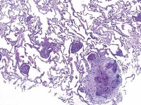
Fig 3 Sarcoidosis. Granulomas involve the pulmonary interstitium (transbronchial biopsy). The adjacent alveolar septa were not inflamed.

Fig 4 Sarcoidosis. A, Pleural involvement with confluent nonnecrotizing granulomas. B, Nonnecrotizing granulomas infiltrate into pleural adipose tissue.
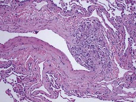
Fig 5 Sarcoidosis. Granulomatous vasculitis characterized by presence of nonnecrotizing granuloma within the vessel wall without causing vascular necrosis.

Fig 6 Sarcoidosis. Granuloma involving the lymphatic vessel wall.

Fig 7 Sarcoidosis. Well-formed granuloma composed of tightly clustered epithelioid histiocytes, multinucleated giant cell, and a few inflammatory cells.

Fig 8 Sarcoidosis. Nonspecific cytoplasmic asteroid inclusion within giant cell.

Fig 9 Sarcoidosis. Hyalinized fibrous tissue replacing portions of granulomas or even entire granulomas; only multinucleated giant cells are present in this treated patient.

Fig 10 Sarcoidosis. Necrotizing granuloma with small area of necrosis present within the granuloma.