
Definition
• Rare condition of lipoproteinaceous components accumulating in alveolar spaces
Pathophysiology
• Autoimmune PAP: neutralizing autoantibodies to granulocyte-macrophage colony-stimulating factor (GM-CSF) resulting in impaired clearance and subsequent accumulation of surfactant proteins
• Secondary PAP: decreased numbers and impaired function of alveolar macrophages due to the underlying chronic disease results in impaired clearance and subsequent accumulation of surfactant proteins
• Hereditary PAP: mutations in the gene encoding the α chain of the GM-CSF receptor (CSF2RA) resulting in reduced binding of GM-CSF, reduced signaling, and reduced GM-CSF–dependent function in alveolar macrophages
Clinical features
Epidemiology
• Autoimmune PAP (formerly idiopathic PAP) is the most common type and occurs in adults
• Secondary PAP most often occurs in adults and is associated with underlying environmental exposures such as silicosis, hematological malignancies, immunodeficiency, or certain infections
• Hereditary PAP is very rare and usually presents in children
• Surfactant disorders (formerly congenital PAP) occur in infants and children and are due to genetic defects of surfactant proteins or proteins involved in surfactant processing and homeostasis; this topic is discussed further in Surfactant Disorders
Presentation
• Adults typically present between 30 to 50 years of age with male to female ratio of 2:1
• Progressive dyspnea is the most common symptom, but patients may also have nonproductive cough, weight loss, fatigue, and fever, especially in secondary PAP
• Some patients may be asymptomatic, with incidental discovery of disease
• May have increased lactate dehydrogenase, polycythemia, or hypergammaglobulinemia, and usually restrictive defect on pulmonary function testing
• Elevated serum anti–GM-CSF titer in patients with autoimmune PAP
• Signs and symptoms of underlying disease in patients with secondary PAP
• Tachypnea, failure to thrive, and increased GM-CSF levels in bronchioalveolar lavage (BAL) fluid in patients with hereditary PAP, although patients may be clinically unrecognized and discovered during familial screening for an affected relative
• High-resolution CT scanning shows homogeneous ground glass opacities with “crazy-paving” pattern, which is suggestive of—but not specific for—PAP
Prognosis and treatment
• Moderate to severely symptomatic autoimmune PAP: whole lung lavage to remove lipoproteinaceous material and anti–GM-CSF antibodies; subcutaneous or aerosolized GM-CSF
• Moderately to severely symptomatic secondary PAP: treatment of underlying disorder or whole lung lavage
• Mildly symptomatic PAP: close monitoring and supplemental oxygen
• Asymptomatic PAP: close monitoring but no specific therapy
• Patients presenting with more severe disease generally have worse prognosis, with increased risk of recurrence or persistent symptoms
• Patients with secondary PAP, especially when associated with a hematological malignancy, tend to have a worse prognosis
• Hereditary PAP is slowly progressive due to some residual GM-CSF–dependent function of alveolar macrophages
Pathology
Histology
• Papanicolaou-stained BAL fluid with 18 or more dense dark-orange globules with sharp green borders
• Histological sections show preserved lung architecture and filling of alveolar spaces by amorphous eosinophilic granular material, which is PAS positive and mucicarmine negative
• Type II pneumocyte hyperplasia, alveolar macrophages, sloughed pneumocytes, and cholesterol clefts may also be present
• Causative particles may be identified in secondary PAP associated with environmental etiologies
• Electron microscopy shows tubular and fused membrane lamellar body–like structures, as well as crystals and cell debris in the airspaces and within alveolar macrophages
Immunopathology (including immunohistochemistry)
• Alveolar eosinophilic material is positive for surfactants A, B, and C
Main differential diagnoses
• Clinical
• Acute respiratory distress syndrome
• Lipoid pneumonia
• Acute interstitial pneumonia
• Drug toxicity or hypersensitivity reaction
• Histological
• Autoimmune PAP and hereditary PAP may look histologically identical to secondary PAP; therefore clinical history, laboratory studies, and stains for microorganisms are important for ruling out secondary PAP
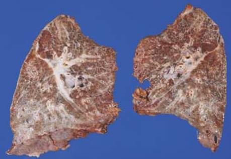
Fig 1 Pulmonary alveolar proteinosis. Grossly, this lung with autoimmune PAP shows dilated airways and cystic spaces filled with glistening material, especially in the lower lobes.
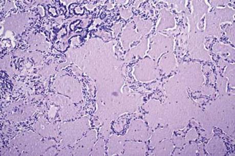
Fig 2 Pulmonary alveolar proteinosis. At low power, the lung architecture is preserved and the alveolar spaces are filled with amorphous eosinophilic granular material.
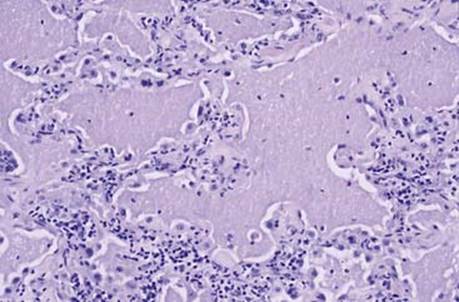
Fig 3 Pulmonary alveolar proteinosis. At high power, hyperplastic type II pneumocytes are seen lining the alveolar spaces. A mild inflammatory infiltrate in the alveolar septa is also present.
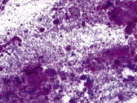
Fig 4 Pulmonary alveolar proteinosis. A 4-year-old with treated acute promyelocytic leukemia developed bilateral diffuse infiltrates. Touch preparation made at the time of frozen section and stained with H&E shows granular material typical of PAP.
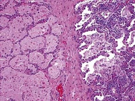
Fig 5 Pulmonary alveolar proteinosis. Intraalveolar proteinaceous material with cholesterol clefts is present. Reactive pneumocytes and interstitial chronic inflammation are seen on the right. Same patient as in Fig 4.
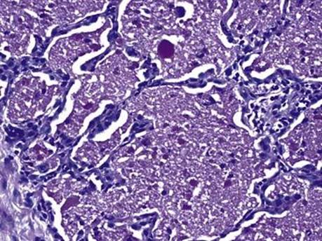
Fig 6 Pulmonary alveolar proteinosis. PAS stain is positive and highlights the granular nature of the intraalveolar accumulation. Same patient as in Fig 4.