
Definition
• Amyloidosis is the extracellular deposition of misfolded proteins in beta pleated sheets
• Primary pulmonary amyloidosis (PPA): amyloid deposition in the respiratory tract without associated systemic amyloidosis. There are two main types of PPA: tracheobronchial amyloidosis (TBA) is the deposition of amyloid limited to the trachea and bronchi, and nodular amyloidosis (NA) is the deposition of balls of amyloid (usually composed of lambda light chain) within the lung parenchyma
• Secondary pulmonary amyloidosis (SPA): amyloid deposition in lung parenchyma and around blood vessels in patient with systemic amyloidosis
Clinical features
Epidemiology and pathogenesis
• NA is rare and can be associated with chronic inflammation seen in Sjögren syndrome, rheumatoid arthritis, systemic lupus erythematosus, tuberculosis, HIV infection, or hematological neoplasms such as multiple myeloma or lymphoplasmacytic lymphoma
• TBA is also rare, occurs in the fifth to sixth decade of life, and can be associated with tracheobronchopathia osteoplastica
Presentation
• Patients with TBA may present with coughing, dyspnea, hemoptysis, pleural effusions, pneumonia, or bronchiectasis
• NA is usually an incidental finding on imaging in asymptomatic patients
Prognosis and treatment
• Patients with TBA are at risk of death secondary to respiratory failure or recurrent pneumonia due to airway obstruction. Treatment may include surgical resection, bronchoscopic resection, or laser ablation
• NA is associated with a benign course. Surgical resection of the nodules is curative as well as diagnostic
Pathology
Gross
• NA: single or multiple nodules measuring 1 to 4 cm; nodules are firm, irregular and yellow to gray
• TBA: diffusely edematous mucosa or nodules protruding into lumen; waxy gray-white cut surface
Histology
• Amorphous eosinophilic material with a foreign body giant cell reaction
• NA: forms a nodule within lung parenchyma
• TBA: deposition limited to submucosa of trachea and bronchi
• NA will have a lymphoplasmacytic infiltrate, usually at the periphery of the nodules
• May have calcification, chondrification, or ossification
• In SPA amorphous eosinophilic material is deposited in alveolar septae and blood vessel walls
Immunopathology/special stains
• Apple green birefringence when Congo red stain is viewed under polarized light
Main differential diagnoses
• NA: hyalinizing granuloma, lymphoma (especially mucosa-associated lymphoid tissue lymphoma) with associated amyloid deposition
• TBA: tracheobronchopathia osteoplastica
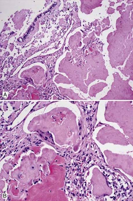
Fig 1 Pulmonary amyloidosis. Amorphous eosinophilic material surrounded by giant cell reaction at low (A) and intermediate (B) powers.

Fig 2 Pulmonary amyloidosis. Congo red stain is positive (i.e., red, with regular light) (A) and displays apple green birefringence of amyloid under polarized light (B).

Fig 3 Pulmonary amyloidosis. SPA: amyloid deposits in blood vessel wall (A) and alveolar wall (B).
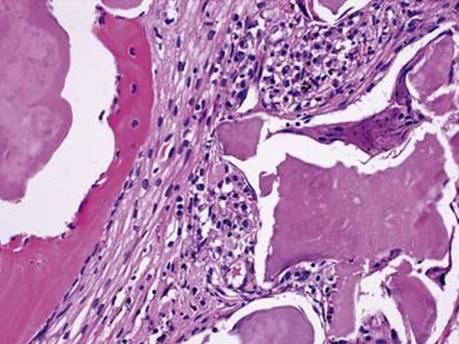
Fig 4 Pulmonary amyloidosis. Amyloid with ossification.