
Definition
• Small, underdeveloped lungs with less than normal weight and fewer than normal alveoli expected for gestational age as determined by lung weight to body weight ratio or radial alveolar count
Clinical features
Epidemiology
• True incidence is unknown but has been noted in less than 10% of neonatal autopsies
• Associated with other fetal anomalies
• Oligohydramnios secondary to renal agenesis or prolonged fetal membrane rupture
• Decreased intrathoracic space secondary to renal cystic disease or diaphragmatic hernia
• Reduced breathing secondary to anencephaly or musculoskeletal disorders
Presentation
• Usually detected on fetal ultrasound
• Severe difficulty in breathing after birth
Prognosis and treatment
• High mortality rate of 70% to 95%
• Lung weight less than 40% of normal is associated with immediate death
• In surviving patients, treatment is supportive
Pathology
Gross
• Small, underdeveloped lungs for gestational age with reduced lung weight to body weight ratio (normal ratio is 0.22)
Histology
• Histologically underdeveloped lungs for gestational age
• Acini are fewer for gestational age, but alveolar and capillary development is unaffected
• There is often an increased amount of connective tissue/mesenchyme giving an immature appearance
• Reduced radial alveolar count
• Draw a line from a terminal bronchiole to the closest septal division or pleural surface. Count the number of intersected alveoli. The mean alveolar count for term infants is 4.4 ± 0.9.
Immunopathology/special stains
• Not contributory
Main differential diagnosis
• Atelectasis: small lungs but weight is normal

Fig 1 Pulmonary hypoplasia. X-ray film of a fetus with pulmonary hypoplasia secondary to diaphragmatic hernia.
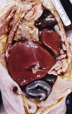
Fig 2 Pulmonary hypoplasia. Gross image of a fetus with pulmonary hypoplasia secondary to diaphragmatic hernia.

Fig 3 Pulmonary hypoplasia. Lungs from the case in Fig 2 are both small; the left lung is smaller. Note that the lungs do not reach the apex of the heart—a good indication of pulmonary hypoplasia.
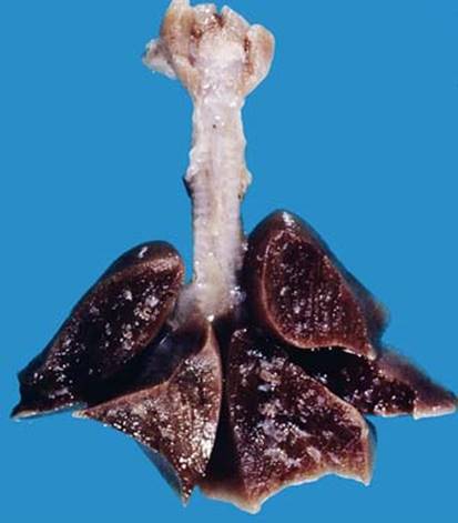
Fig 4 Pulmonary hypoplasia. Cut surface of hypoplastic lungs.

Fig 5 Pulmonary hypoplasia. Low (A) and high (B and C) powers show small lobules with fewer acini than would be expected in this term baby with renal agenesis.