
Definition
• Developmental anomaly of the lower respiratory tract
• Previously known as congenital cystic adenomatoid malformation (CCAM)
Pathogenesis
• Abnormalities of lung branching morphogenesis thought to occur at different stages and levels of lung development and classified accordingly into five types
Clinical features
Epidemiology
• Most common congenital lung lesion, 1 per 5000 to 35,000 live births
• Most occur sporadically (see Table 1 in the Appendix)
Presentation
• Affected patients may present with respiratory distress in the newborn period or may remain asymptomatic until later in life (see Table 1 in the Appendix)
• Many cases are now detected by routine prenatal ultrasound examination
Prognosis and treatment
• Spontaneous regression can occur but is infrequent
• Immediate surgical resection is indicated in symptomatic patients
• Surgery can be delayed for a few years in asymptomatic patients; there are rare case reports of bronchioloalveolar carcinoma arising in CPAM in the second and third decades
Pathology
• See Table 1 in the Appendix (gross and microscopic)
Immunopathology/special stains
• Alveolar type II cells lining CPAM4 can be highlighted by immunohistochemical analysis for TTF-1, cytokeratins, or surfactant
Main differential diagnoses
• Resolution of an abscess/necrotic process: can resolve forming cysts with lining similar to CPAM. Chronic inflammation and fibrosis are present. These cannot be definitively distinguished from CPAM with superimposed chronic inflammation unless clinical history is clear
• Cystic pleuropulmonary blastoma: cambium layer beneath the epithelial lining of the cysts contains desmin and myogenin-positive malignant cells (rhabdomyoblasts)
• Bronchogenic cyst: seen in older children, not connected to tracheobronchial tree, filled with acellular material (not air), and does not have surrounding smaller cysts
• Persistent interstitial pulmonary emphysema: cystic spaces have foreign body giant cells and no epithelial lining
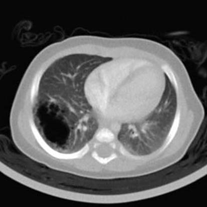
Fig 1 Congenital pulmonary airway malformation. This computed tomography scan shows a 3-cm central cyst with surrounding smaller cysts in an infant with CPAM type 1.
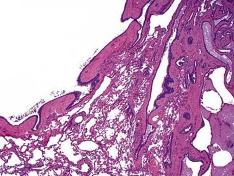
Fig 2 Congenital pulmonary airway malformation. The main cyst and adjacent smaller cysts have a thick fibrous wall in CPAM type 1.
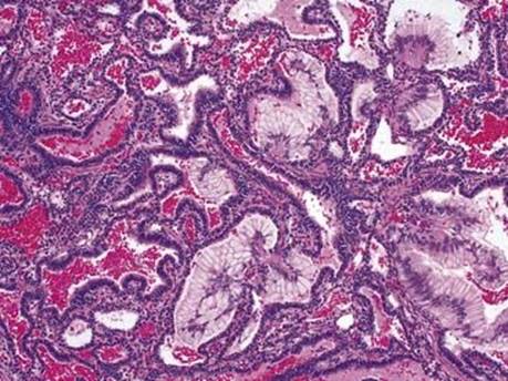
Fig 3 Congenital pulmonary airway malformation. Multiple clusters of mucogenic cells are present in this CPAM type 1.
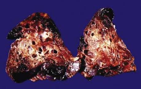
Fig 4 Congenital pulmonary airway malformation. This cut surface of the lung shows type 2 CPAM with multiple small cysts and solid areas blending with normal lung.
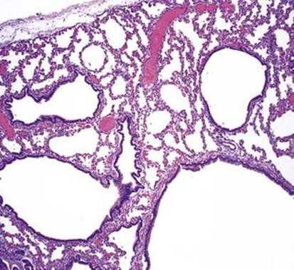
Fig 5 Congenital pulmonary airway malformation. Type 2 CPAM has numerous bronchiole-like spaces extending into peripheral lung tissue.
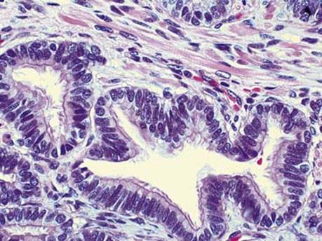
Fig 6 Congenital pulmonary airway malformation. This type 2 CPAM has striated muscle cells in the wall.
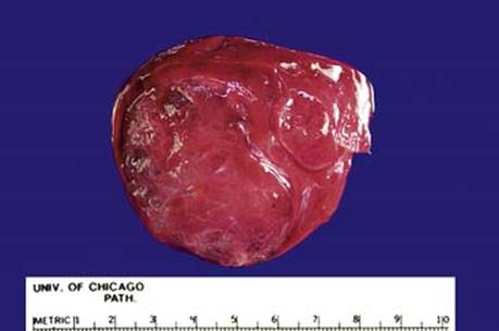
Fig 7 Cut surface of lung resection with CPAM type 3 shows no grossly visible cysts.
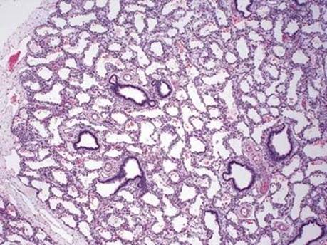
Fig 8 Congenital pulmonary airway malformation. This type 3 CPAM resembles the canalicular stage of lung development.
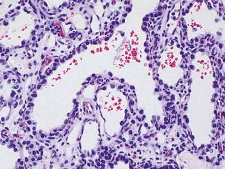
Fig 9 Congenital pulmonary airway malformation. High power of Fig 8 shows that the spaces are lined by low cuboidal cells.
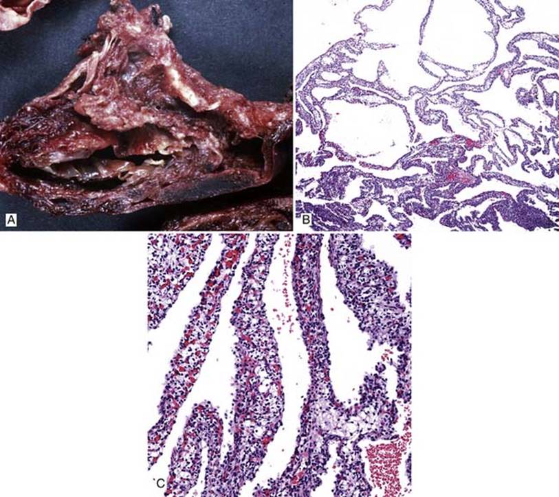
Fig 10 Congenital pulmonary airway malformation. Type 4 CPAM: A, gross image showing large cysts; B, low power showing thin-walled cysts; and C, high power showing cyst walls lined by pneumocytes and containing histiocytic cells with clear cytoplasm, reminiscent of pulmonary interstitial glycogenosis.
(Courtesy of Dr. J.T. Stocker, Bethesda, Md.)