
When assessing a lung biopsy specimen from a patient clinically thought to have an infectious process, it is important to consider the immune status of the patient, as this has a bearing on the differential diagnosis. For example, in a transplant patient, opportunistic infections, such as cytomegalovirus (CMV) or Pneumocystis jiroveci would be high on the differential diagnosis list, whereas in a child, respiratory syncytial virus (RSV) or adenovirus would be a strong consideration. In those with alcoholism or elderly patients with pneumonia, one must think of an aspiration pneumonia involving mixed oral flora, while an otherwise healthy adult with community-acquired pneumonia would have findings caused by a single organism. There are a few important points to keep in mind regarding immunocompromised patients and opportunistic infections. First, common infectious agents are common and still must be considered in immunocompromised patients. Second, an opportunistic infection can be found in patients without any prior history as the first significant manifestation of an underlying disease. Finally, in immunocompromised patients, the finding of one opportunistic infection should spur, not deter, the search for others, as coinfection with multiple organisms is not uncommon.
Please see Tables 4, 5, and 6 in the Appendix for a summary of key features of bacterial, viral, and fungal pneumonias, respectively.
Definition
• Pulmonary diseases in patients with AIDS, not directly related to the immunosuppressive effects of HIV
• Noninfectious pulmonary involvement in patients with HIV can be broadly classified as follows:
• Lymphocytic interstitial pneumonia
• Malignancy
• Vascular disorders
• Therapy-related changes
Clinical features
Epidemiology
• Noninfectious pulmonary complications in HIV patients are on the rise because of improvement in overall survival with the advent of highly active antiretroviral therapy (HAART)
Presentation
• Nonspecific symptoms include weight loss and a fever; specific symptoms like shortness of breath, chest pain, and a cough may also be present
• Lymphocytic interstitial pneumonia is frequent in patients with altered immune states including AIDS and other autoimmune disorders and may be the presenting feature, especially in children
• Pulmonary arterial hypertension (PAH) associated with HIV is an infrequent (0.5% of cases) but serious complication associated with significant morbidity and mortality. Chest radiography shows cardiomegaly with right ventricular hypertrophy and a prominent pulmonary artery. There is increased vascular resistance with increase in diastolic and systolic arterial pressures
• Imaging studies, blood cultures, and laboratory work-up are helpful in ruling out coexisting infectious agents
Pathology
Histology
• Lymphoid interstitial pneumonia (LIP): alveolar walls are thickened because of infiltration by a polymorphous population composed of small lymphocytes (mainly T cells) and plasma cells. Poorly formed granulomas and aggregates of histiocytes may also be seen. The involvement is typically bilateral and interstitial (refer to the section on LIP for further details)
• Pulmonary hypertension: plexiform arteriopathy with dilated vascular channels filled with intraluminal capillary channels resembling an organized thrombus (refer to section on pulmonary hypertension for further details)
• Malignancies include lung cancer, Kaposi sarcoma, lymphoproliferative disorders
• Lung cancers in HIV-positive patients do not differ from those in HIV-negative patients with lung cancer in terms of histology and overall survival. However, the HIV-positive patients have an earlier onset of the disease
Main differential diagnoses
• Lymphoid lung infiltrates may also be seen in primary pulmonary marginal zone lymphomas, which are generally monoclonal, unifocal, mass forming, Bcl-2 positive and Bcl-6 negative
• Infections: appropriate special stains

Fig 1 Noninfectious pulmonary lesions in HIV/AIDS patients. Low- (A) and intermediate- (B) power images of a lung biopsy from an AIDS patient seen with LIP. There is a varying amount of diffuse lymphoplasmacytic infiltrate in the interstitium.
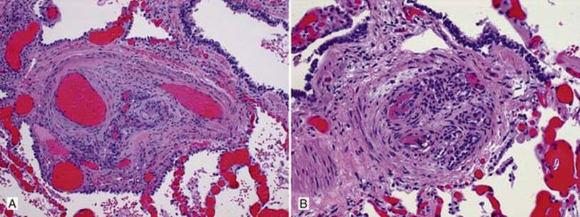
Fig 2 Noninfectious pulmonary lesions in HIV/AIDS patients. Plexiform lesions in an AIDS patient with multiple intraarterial lumina resembling recanalized thrombus (A) and multiple endothelial lined intravascular spaces with fibrin thrombi (B).
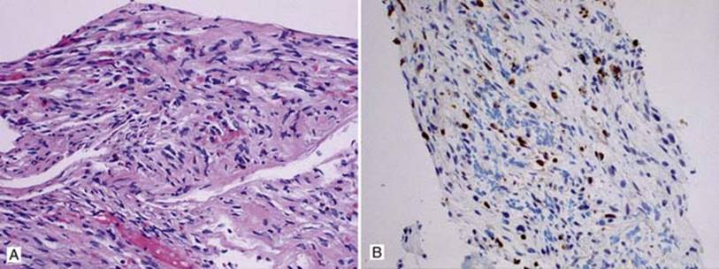
Fig 3 Noninfectious pulmonary lesions in HIV/AIDS patients. Needle biopsy of the lung from an AIDS patient. A, There are spindle cells, red blood cells and hemosiderin; however, the amount of infiltrate is scant. B, Positive immunohistochemical testing for HHV-8 is very helpful in diagnosing Kaposi sarcoma. Note that hemosiderin stains darkly and should not be mistaken for positive cells, which have smooth round-to-oval nuclear outlines.